Perspective on the Pandemic
By Jeff Harris | Ron Paul Institute | September 17, 2021
According to the CDC, as of April 26, 2021, 3,427,321 people died from all causes in the United States in 2020. In comparison, 2,854,838 people died from all causes in the US in 2019 of which the CDC says 345,323 died from Covid-19. Trouble is we know a large percentage of these deaths were “with” Covid, not necessarily “from” Covid.
Yes, the total number of deaths was up in 2020 but the fact is we really don’t know exactly how many were attributable to Covid alone. Some estimates suggest the actual mortality from Covid is in the seven percent range of the CDC estimates, suggesting that 93 percent of the deaths were due to something else.
We do know the CDC says that Covid-19 is lethal to .026 percent of those infected meaning 99.74 percent survive. So if there were 345,323 deaths in 2020, allegedly due to Covid-19 that would mean only about 13 Million people were actually infected with Covid? But that doesn’t square with a recent report from the National Institute of Health (NIH) published September 7th, 2021, referencing a Nature magazine article that estimated the actual number of Covid-19 infections in 2020 was over 100 Million?
Using the CDC’s data we can calculate that 345,323 deaths would have meant 13 Million were infected assuming the CDC’s .026 percent mortality rate is accurate. If the Nature article is accurate that means Covid-19 is actually much less lethal than the CDC says. Somebody is lying through their teeth.
Aging Americans
It’s no secret the US has a rapidly aging population. According to the US Census Bureau there were over 54 million people age 65 and over in 2019. Of these more than 13 million are over age 80. According to the CDC the average life expectancy for US males is 76 and for females 81.
So virtually the entire population age 80+ is beyond their normal life expectancy. And according to the CDC 85 percent of people over age 65 have at least one of the following six chronic health conditions: diabetes, cardiovascular disease, chronic obstructive pulmonary disease, asthma, cancer, or arthritis. And 56 percent (over half) have at least two chronic health conditions.
Obviously as one ages the probability of suffering from chronic health ailments goes up. Interestingly, the CDC does not list obesity as a chronic health ailment. According to The National Conference of State Legislatures obesity is a big deal. In their September 2014 report they said:
Obesity, a common and costly health issue that increases risk for heart disease, type 2 diabetes, and cancer, affects more than one-third of adults and 17 percent of youth in the United States. By the numbers, 78 million adults and 12 million children are obese—figures many regard as an epidemic…
So way back in 2014 it was obvious the US was suffering from an obesity “epidemic” according to this report. According to the CDC nearly 25 percent of Americans over age 65 are obese and as we saw above over half of those people have at least two chronic health issues.
The Obesity ‘Epidemic’
So if one out of four Americans over age 65 is obese, with all the serious health consequences that entails AND half of them have at least two chronic health issues as well, that would suggest it wouldn’t take much to push them over the edge to meet their maker. All it would take is a bad flu bug, a bout of pneumonia, or a case of bronchitis combined with existing health problems for death to occur.
How many of these deaths could have been prevented if timely (and as we now know, effective) treatments had been administered like Ivermectin and Hydroxychloroquine? Many doctors who were actually treating patients with these protocols reported dramatic improvement in 36-72 hours. But instead our medical “experts” literally outlawed these treatments and cancelled any doctor who spoke publicly about their effectiveness?
This Isn’t about health care
An additional 345,323 deaths in 2020 works out to about .65 percent of the 65 and over population in the US. Is that really a dramatic increase in deaths when proven effective treatments were purposely withheld from sick patients by medical professionals? And what about all the people who were refused routine medical care because of the hospitals being overwhelmed with patients?
But somehow these same hospitals that were allegedly “overwhelmed” found time for their staff to perform elaborately choreographed dance routines that were uploaded to Youtube and TikTok.
So far I’ve focused on physical ailments but this “pandemic” has been as much a psychological attack on the people as a physical one. Here’s why this matters: In the Holy Bible Proverbs 23:7 says, “As a man thinks in his heart so is he.” In this case the “heart” is the subconscious mind.
According to the Cleveland Clinic, five-to-seven percent of the adult population is susceptible to what is known as “somatic” disorder or psychosomatic disorder. They emphasize that somatic patients have weakened immune systems due to their condition making them more susceptible to illness. Somatic illness is defined as:
Somatic symptom disorder is a disorder in which individuals feel excessively distressed about their health and also have abnormal thoughts, feelings, and behaviors in response to their symptoms. There are different subtypes of the disorder based on the patient’s complaint. The disorder causes a disruption in the patient’s normal functioning and quality of life.
According to the latest Census data there are approximately 250 million adults in the US So if we take the lower five percent estimate that would suggest there are over 12 million adults predisposed to somatic illness. How do you think these people have been affected by the constant barrage of fear porn being broadcast 24/7 by all types of media?
Mandatory face masks everywhere, social distancing signs plastered on the floor of every store, hand sanitizer everywhere, Plexiglas shields at the bank, restaurants closed down, churches closed, neighbors committing suicide and no access to needed routine medical care. And being told there is NO treatment available until a vaccine arrives. Then discovering the vaccine doesn’t work very well and may be worse than the virus! I suspect some somatic patients have literally died from constant, grinding fear morphing into abject terror.
What has been done by our elected officials and their medical “experts” is the greatest crime against the people that has ever been perpetrated by any criminal gang in the history of the world! And they are doing it under the “authority” of the state which they claim is legal.
It is not and we must resist with every fiber of our being right now or there will be no country worth having for our children and grandchildren.
Surgeon: Jabbing 12 To 15 Year-Olds Is “Dangerous”
By Richie Allen | September 20, 2021
Retired orthopaedic surgeon Professor John Fairclough claimed this morning that jabbing 12 to 15 year-olds is dangerous. He also criticised the government for overruling the Joint Committee on Vaccination and Immunisation (JCVI).
Speaking to Talk Radio’s Julia Hartley-Brewer, Professor Fairclough said:
“I think this is probably… I’ve been in medicine now for five decades. I think this is probably one of the most dangerous efforts that we’ve had in medicine because what we’re doing is we’re in fact overturning the position of medical individuals for political purposes.”
On the issue of informed consent, Fairclough said that it’s unlikely children understand the risks associated with receiving a jab, when SAGE (Scientific Advisory Group for Emergencies) members themselves don’t seem to know. Fairclough said:
“Consent is that you are aware of what the consequences of a medical action is. Recently, one of the members of SAGE chose to go on a radio station and be interviewed with three or four young children and actually wasn’t able to give the information which is on the government website and had to Google it.
He also got the rates of inflammation of the heart incorrect and the published international data. So if he cannot actually be aware of the consent issues, then how can a 12 year-old?”
Fairclough was referring to England’s deputy chief medical officer Jonathan Van Tam. While fielding questions from youngsters on BBC News recently, Van Tam was caught with his trousers well and truly down when wrongly downplaying the threat of myocarditis. The jellyfish in the presenters chair never called him on it.
Rather than challenge the witchdoctors who currently advise the UK government, the media has deified them. Earlier this year, speaking on BBC Radio 5 Live, presenter Nicky Campbell said: “We all have our favourite scientist now. Isn’t JVT (Van Tam) great?”
I don’t know which is worse, the liars in Bill Gates pocket, or the whores in the media.
APOE-4: The Clue to Why Low Fat Diet and Statins may Cause Alzheimer’s
By Dr. Stephanie Seneff | December 15, 2009
Abstract
Alzheimer’s is a devastating disease whose incidence is clearly on the rise in America. Fortunately, a significant number of research dollars are currently being spent to try to understand what causes Alzheimer’s. ApoE-4, a particular allele of the apolipoprotein apoE, is a known risk factor. Since apoE plays a critical role in the transport of cholesterol and fats to the brain, it can be hypothesized that insufficient fat and cholesterol in the brain play a critical role in the disease process. In a remarkable recent study, it was found that Alzheimer’s patients have only 1/6 of the concentration of free fatty acids in the cerebrospinal fluid compared to individuals without Alzheimer’s. In parallel, it is becoming very clear that cholesterol is pervasive in the brain, and that it plays a critical role both in nerve transport in the synapse and in maintaining the health of the myelin sheath coating nerve fibers. An extremely high-fat (ketogenic) diet has been found to improve cognitive ability in Alzheimer’s patients. These and other observations described below lead me to conclude that both a low-fat diet and statin drug treatment increase susceptibility to Alzheimer’s.
1. Introduction
Alzheimer’s is a devastating disease that takes away the mind bit by bit over a period of decades. It begins as odd memory gaps but then steadily erodes your life to the point where around-the-clock care is the only option. With severe Alzheimer’s, you can easily wander off and get lost, and may not even recognize your own daughter. Alzheimer’s was a little known disease before 1960, but today it threatens to completely derail the health system in the United States.
Currently, over 5 million people in America have Alzheimer’s. On average, a person over 65 with Alzheimer’s costs three times as much for health care as one without Alzheimer’s. More alarmingly, the incidence of Alzheimer’s is on the rise. Dr. Murray Waldman has studied epidemiological data comparing Alzheimer’s with femur fractures, looking back over the last fifty years [52]. Alarmingly, he has found that, while the incidence of femur fractures (another condition which typically increases with age) has gone up only at a linear rate, the increase in the incidence of Alzheimer’s has gone up exponentially, between 1960 and 2010 Alzheimer’s Epidemic [15]. Just between 2000 and 2006, US Alzheimer’s deaths rose by 47%, while, by comparison, deaths from heart disease, breast cancer, prostate cancer, and stroke combined decreased by 11%. This increase goes far beyond people living longer: for people 85 and older, the percentage who died from Alzheimer’s rose by 30% between 2000 and 2005 [2]. Finally, it’s likely these are under-estimates, as many people suffering with Alzheimer’s ultimately die of something else. You likely have a close friend or relative who is suffering from Alzheimer’s.
Something in our current lifestyle is increasing the likelihood that we will succumb to Alzheimer’s. My belief is that two major contributors are our current obsession with low-fat diet, combined with the ever expanding use of statin drugs. I have argued elsewhere that low-fat diet may be a major factor in the alarming increase in autism and adhd in children. I have also argued that the obesity epidemic and the associated metabolic syndrome can be traced to excessive low-fat diet. Statins are likely contributing to an increase in many serious health issues besides Alzheimer’s, such as sepsis, heart failure, fetal damage, and cancer, as I have argued here. I believe the trends will only get worse in the future, unless we substantially alter our current view of “healthy living.”
The ideas developed in this essay are the result of extensive on-line research I conducted to try to understand the process by which Alzheimer’s develops. Fortunately, a great deal of research money is currently being spent on Alzheimer’s, but a clearly articulated cause is still elusive. However, many exciting leads are fresh off the press, and the puzzle pieces are beginning to assemble themselves into a coherent story. Researchers are only recently discovering that both fat and cholesterol are severly deficient in the Alzheimer’s brain. It turns out that fat and cholesterol are both vital nutrients in the brain. The brain contains only 2% of the body’s mass, but 25% of the total cholesterol. Cholesterol is essential both in transmitting nerve signals and in fighting off infections.
A crucial piece of the puzzle is a genetic marker that predisposes people to Alzheimer’s, termed “apoE-4.” ApoE plays a central role in the transport of fats and cholesterol. There are currently five known distinct variants of apoE (properly termed “alleles”), with the ones labelled “2”, “3” and “4” being the most prevalent. ApoE-2 has been shown to afford some protection against Alzheimer’s; apoE-3 is the most common “default” allele, and apoE-4, present in 13-15% of the population, is the allele that is associated with increased risk to Alzheimer’s. A person with apoE-4 allele inherited from both their mother and their father has up to a twenty-fold increased likelihood of developing Alzheimer’s disease. However, only about 5% of the people with Alzheimer’s actually have the apoE-4 allele, so clearly there is something else going on for the rest of them. Nonetheless, understanding apoE’s many roles in the body was a key step leading to my proposed low fat/statin theory.
2. Background: Brain Biology 101
Although I have tried to write this essay in a way that is accessible to the non-expert, it will still be helpful to first familiarize you with basic knowledge of the structure of the brain and the roles played by different cell types within the brain.
At the simplest level, the brain can be characterized as consisting of two major components: the gray matter and the white matter. The gray matter comprises the bodies of the neurons, including the cell nucleus, and the white matter contains the myriad of “wires” that connect each neuron to every other neuron it communicates with. The wires are known as “axons” and they can be quite long, connecting, for example, neurons in the frontal cortex (above the eyes) with other neurons deep in the interior of the brain concerned with memory and movement. The axons will figure prominently in the discussions below, because they are coated with a fatty substance called the myelin sheath, and this insulating layer is known to be defective in Alzheimer’s. Neurons pick up signals transmitted through the axons at junctures known as synapses. Here the message needs to be transmitted from one neuron to another one, and various neurotransmitters such as dopamine and GABA exert excitatory or inhibitory influences on signal strength. In adidtion to a single axon, neurons typically have several much shorter nerve fibers called dendrites, whose job is to receive incoming signals from diverse sources. At a given point in time, signals received from multiple sources are integrated in the cell body and a decision is made as to whether the accumulated signal strength is above threshold, in which case the neuron responds by firing a sequence of electrical pulses, which are then transmitted through the axon to a possibly distant destination.
In addition to the neurons, the brain also contains a large number of “helper” cells called glial cells, which are concerned with the care and feeding of neurons. Three principle types of glial cells will play a role in our later discussion: the microglia, the astrocytes, and the oligodendrocytes. Microglia are the equivalent of white blood cells in the rest of the body. They are concerned with fighting off infective agents such as bacteria and viruses, and they also monitor neuron health, making life-and-death decisions: programming a particular neuron for apoptosis (intentional self-destruction) if it appears to be malfunctioning beyond hope of recovery, or is infected with an organism that is too dangerous to let flourish.
The astrocytes figure very prominently in our story below. They nestle up against the neurons and are responsible for assuring an adequate supply of nutrients. Studies on neuron cultures from rodent central nervous systems have shown that neurons depend upon astrocytes for their supply of cholesterol [40]. Neurons critically need cholesterol, both in the synapse [50] and in the myelin sheath [45], in order to successfully transmit their signals, and also as a first line of defense against invasive microbes. Cholesterol is so important to the brain that astrocytes are able to synthesize it from basic ingredients, a skill not found in most cell types. They also supply the neurons with fatty acids, and they are able to take in short chain fatty acids and combine them to form the longer-chain types of fatty acids that are especially prominent in the brain [7][24][36], and then deliver them to neighboring neurons and to the cerebrospinal fluid.
The third type of glial cell is the oligodendrocyte. These cells specialize in making sure the myelin sheath is healthy. Oligodentrocytes synthesize a special sulfur-containing fatty acid, known as sulfatide, from other fatty acids supplied to them by the cerebrospinal fluid [9]. Sulfatide has been shown to be essential for the maintenance of the myelin sheath. Children born with a defect in the ability to metabolize sulfatide suffer from progressive demyelination, and rapid loss of motor and cognitive functions, resulting in an early death before the age of 5 [29]. Depletion in sulfatide is a well-known characterization of Alzheimer’s, even in early stages before it has been manifested as cognitive decline [18]. And ApoE has been shown to play a crucial role in the maintenance of sulfatide [19]. Throughout a person’s life, the myelin sheath has to be constantly maintained and repaired. This is something that researchers are only beginning to appreciate, but two related properties of Alzheimer’s are poor quality myelin sheath alongside a drastically reduced concentration of fatty acids and cholesterol in the cerebrospinal fluid [38].
3. Cholesterol and Lipid Management
In addition to some knowledge about the brain, you will also need to know something about the processes that deliver fats and cholesterol to all the tissues of the body, with a special focus on the brain. Most cell types can use either fats or glucose (a simple sugar derived from carbohydrates) as a fuel source to satisfy their energy needs. However, the brain is the one huge exception to this rule. All cells in the brain, both the neurons and the glial cells, are unable to utilize fats for fuel. This is likely because fats are too precious to the brain. The myelin sheath requires a constant supply of high quality fat to insulate and protect the enclosed axons. Since the brain needs its fats to survive long-term, it is paramount to protect them from oxidation (by exposure to oxygen) and from attack by invasive microbes.
Fats come in all kinds of shapes and sizes. One dimension is the degree of saturation, which concerns how many double bonds they possess, with saturated fats possessing none, monounsaturated fats having only one, and polyunsaturated fats having two or more. Oxygen breaks the double bond and leaves the fat oxidized, which is problematic for the brain. Polyunsaturated fats are thus the most vulnerable to oxygen exposure, because of multiple double bonds.
Fats are digested in the intestine and released into the blood stream in the form of a relatively large ball with a protective protein coat, called a chylomicron. The chylomicron can directly provide fuel to many cell types, but it may also be sent to the liver where the contained fats are sorted out and redistributed into much smaller particles, which also contain substantial amounts of cholesterol. These particles are called “lipoproteins,” (henceforth, LPP’s) because they contain protein in the spherical shell and lipids (fats) in the interior. If you’ve had your cholesterol measured, you’ve probably heard of LDL (low density LPP) and HDL (high density LPP). If you think these are two different kinds of cholesterol, you would be mistaken. They are just two different kinds of containers for cholesterol and fats that serve different roles in the body. There are actually several other LPP’s, for example, VLDL (very-low) and IDL (intermediate), as shown in the accompanying diagram.  In this essay I will refer to these collectively as the XDL’s. As if this weren’t confusing enough, there is also another unique XDL that is found only in the cerebrospinal fluid, that supplies the nutritional needs of the brain and nervous system. This one doesn’t seem to have a name yet, but I will call it “B-HDL,” because it is like HDL in terms of its size, and “B” is for “brain [13]”
An important point about all the XDL’s is that they contain distinctly different compositions, and each is targeted (programmed) for specific tissues. A set of proteins called “apolipoproteins” or, equivalently, “apoproteins” (“apo’s” for short) figure strongly in controlling who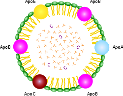 gets what. As you can see from the schematic of the chylomicron shown at the right, it contains a rainbow of different apo’s for every conceivable application. But the XDL’s are far more specific, with HDL containing “A,” LDL containing “B,” VLDL containing “B” and “C,” and IDL containing only “E.” The apo’s have special binding properties that allow the lipid contents to be transported across cell membranes so that the cell can gain access to the fats and choleseterol contained inside.
The only apo that is of concern to us in the context of this essay is apoE. ApoE is very important to our story because of its known link with Alzheimer’s disease. ApoE is a protein, i.e., sequence of amino acids, and its specific composition is dictated by a corresponding DNA sequence on a protein-coding gene. Certain alterations in the DNA code lead to defects in the ability of the transcribed protein to perform its biological roles. ApoE-4, the allele associated with increased risk to Alzheimer’s, is presumably unable to perform its tasks as efficiently as the other alleles. By understanding what apoE does, we can better infer how the consequences of doing it poorly might impact the brain, and then observe experimentally whether the features of the Alzheimer’s brain are consistent with the roles played by apoE.
A strong clue about apoE’s roles can be deduced from where it is found. As I mentioned above, it is the only apo in both B-HDL in the cerebrospinal fluid and IDL in the blood serum. Only selected cell types can synthesize it, the two most significant of which for our purposes are the liver and the astrocytes in the brain. Thus the astrocytes provide the linkage between the blood and the cerebrospinal fluid. They can usher lipids and cholesterol across the blood-brain barrier, via the special key which is apoE.
It turns out that, although apoE is not found in LDL, it does bind to LDL, and this means that astrocytes can unlock the key to LDL in the same way that they can gain access to IDL, and hence the cholesterol and fatty acid contents of LDL are accessible to astrocytes as well, as long as apoE is functioning properly. The astrocytes reshape and repackage the lipids and release them into the cerebospinal fluid, both as B-HDL and simply as free fatty acids, available for uptake by all parts of the brain and nervous system [13].
One of the critical reshaping steps is to convert the fats into types that are more attractive to the brain. To understand this process you need to know about another dimension of fats besides their degree of saturation, which is their total length. Fats have a chain of linked carbon atoms as their spine, and the total number of carbons in a particular fat characterizes it as short, medium length, or long. The brain works best when the constituent fats are long, and, indeed, the astrocytes are able to take in short chain fats and reorganize them to make longer chain fats [24].
A final dimension of fats that plays a role is where the first double bond is located in a polyunsaturated fat, which distinguishes omega-3 from omega-6 fats (position 3; position 6). Omega-3 fats are very common in the brain. Certain ones of the omega-3 and omega-6 fats are essential fatty acids, in that the human body is unable to synthesize them, and therefore depends upon their supply from the diet. This is why it is claimed that fish “makes you smart”: because cold water fish is the best source of essential omega-3 fats.
Now I want to return to the subject of the XDL’s. It is a dangerous journey from the liver to the brain, as both oxygen and microbes are found in abundance in the blood stream. The XDL’s protective shell contains both LPP’s andunesterified cholesterol, as well as the signature apo that controls which cells can receive the contents, as shown in the accompanying schematic.  The internal contents are esterified cholesterol and fatty acids, along with certain antioxidants that are conveniently being transported to the cells packaged in the same cargo ship. Esterification is a technique to render the fats and cholesterol inert, which helps protect them from oxidation [51]. Having the antioxidants (such as vitamin E and Coenzyme Q10) along for the ride is also convenient, as they too protect against oxidation. The cholesterol contained in the shell, however, is intentionally not esterified, which means that it is active. One of its roles there is to guard against invasive bacteria and viruses [55]. Cholesterol is the first line of defense against these microbes, as it will alert the white blood cells to attack whenever it encounters dangerous pathogens. It has also been proposed that the cholesterol in the XDL’s shell itself acts as an antioxidant [48].
HDL’s are mostly depleted of the lipid and cholesterol content, and they are tasked with returning the empty shell back to the liver. Once there, cholesterol will be recommissioned to enter the digestive system as part of the bile, which is produced by the gall bladder to help digest ingested fats. But the body is very careful to conserve cholesterol, so that 90% of it will be recycled from the gut back into the blood stream, contained in the chylomicron that began our story about fats.
In summary, the management of the distribution of fats and cholesterol to the cells of the body is a complex process, carefully orchestrated to assure that they will have a safe journey to their destination. Dangers lurk in the blood stream, mostly in the form of oxygen and invasive microbes. The body considers cholesterol to be precious cargo, and it is very careful to conserve it, by recycling it from the gut back to the liver, to be appropriately distributed among the XDL’s that will deliver both cholesterol and fats to the tissues that depend upon them, most especially the brain and nervous system.
4. The Relationship between Cholesterol and Alzheimer’s
Through retrospective studies, the statin industry has been very successful at the game of pretending that benefits derived from high cholesterol are actually due to statins, as I have described at length in an essay on the relationship between statins and fetal damage, sepsis, cancer, and heart failure. In the case of Alzheimer’s, they are playing this game in reverse: they are blaming cholesterol for a very serious problem that I believe is actually caused by statins.
The statin industry has looked long and hard for evidence that high cholesterol might be a risk factor for Alzheimer’s. They examined cholesterol levels for men and women of all ages between 50 and 100, looking back 30 or more years if necesssary, to see if there was ever a correlation between high cholesterol and Alzheimer’s. They found only one statistically significant relationship: men who had had high cholesterol in their 50’s had an increased susceptibility to Alzheimer’s much later in life [3].
The statin industry has jumped on this opportunity to imply that high cholesterol might cause Alzheimer’s, and, indeed, they have been very fortunate in that reporters have taken the bait and are promoting the idea that, if high cholesterol many years ago is linked to Alzheimer’s, then statins might protect from Alzheimer’s. Fortunately, there exist lengthy web pages (Cholesterol Doesn’t Cause Alzheimer’s) that have documented the long list of reasons why this idea is absurd.
Men who have high cholesterol in their 50’s are the poster child for statin treatment: all of the studies that have shown a benefit for statins in terms of reducing the number of minor heart attacks involved men in their 50’s. High cholesterol is positively correlated with longevity in people over 85 years old [54], and has been shown to be associated with better memory function [53] and reduced dementia [35]. The converse is also true: a correlation between falling cholesterol levels and Alzheimer’s [39]. As will be discussed further later, people with Alzheimer’s also have reduced levels of B-HDL, as well as sharply reduced levels of fatty acids, in the cerbrospinal fluid, i.e, impoverished supply of cholesterol and fats to the myelin sheath [38]. As we saw earlier, fatty acid supply is essential as building blocks for the sulfatide that is synthesized by oligodendrocytes to keep the myelin sheath healthy [29].
The obvious study that needs to be done is to bin the men who had high cholesterol in their 50’s into three groups: those who never took statins, those who took smaller doses for shorter times, and those who took larger doses for longer times. Such a study would not be hard to do; in fact, I suspect something like it has already been done. But you’ll never hear about it because the statin industry has buried the results.
In a very long term retrospective cohort study of members of the Permanente Medical Care Program in northern California, researchers looked at cholesterol data that were obtained between 1964 and 1973 [46]. They studied nearly ten thousand people who had remained members of that health plan in 1994, upon the release of computerized outpatient diagnoses of dementia (both Alzheimer’s and vascular dementia). The subjects were between 40 and 45 years old when the cholesterol data were collected.
The researchers found a barely statistically significant result that people who were diagnosed with Alzheimer’s had higher cholesterol in their 50’s than the control group. The mean value for the Alzheimer’s patients was 228.5, as against 224.1 for the controls.
The question that everybody ought to be asking is: for the Alzheimer’s group, how did the people who later took statins stack up against the people who didn’t? In extreme understatement, the authors offhandedly remark in the middle of a paragraph: “Information on lipid-lowering treatments, which have been suggested to decrease dementia risk [31], was not available for this study.” You can be sure that, if there was any inkling that the statins might have helped, these researchers would have been allowed access to those data.
The article they refer to for support, reference [19] in [46] (which is reference [44] here) was very weak. The abstract for that article is repeated in full here in the Appendix. But the concluding sentence sums it up well: “A more than a modest role for statins in preventing AD [Alzheimer’s Disease] seems unlikely.” This is the best they can come up with to defend the position that statins might protect from Alzheimer’s.
An intuitive explanation for why high cholesterol at an early age might be correlated with Alzheimer’s risk has to do with apoE-4. People with that allele are known to have high cholesterol early in life [39], and I believe this is a protective strategy on the part of the body. The apoE-4 allele is likely defective in the task of importing cholesterol into the astrocytes, and therefore an increase in the bioavailability of cholesterol in blood serum would help to offset this deficit. Taking a statin would be the last thing a person in that situation would want to do.
5. Do Statins Cause Alzheimer’s?
There is a clear reason why statins would promote Alzheimer’s. They cripple the liver’s ability to synthesize cholesterol, and as a consequence the level of LDL in the blood plummets. Cholesterol plays a crucial role in the brain, both in terms of enabling signal transport across the synapse [50] and in terms of encouraging the growth of neurons through healthy development of the myelin sheath [45]. Nonetheless, the statin industry proudly boasts that statins are effective at interfering with cholesterol production in the brain [31][47] as well as in the liver.
Yeon-Kyun Shin is an expert on the physical mechanism of cholesterol in the synapse to promote transmission of neural messages, and one of the authors of [50] referenced earlier. In an interview by a Science Daily reporter, Shin said: “If you deprive cholesterol from the brain, then you directly affect the machinery that triggers the release of neurotransmitters. Neurotransmitters affect the data-processing and memory functions. In other words — how smart you are and how well you remember things.”
A recent review of two large population-based double-blind placebo-controlled studies of statin medications in individuals at risk for dementia and Alzheimer disease showed that statins are not protective against Alzheimer’s [34]. The lead author of the study, Bernadette McGuinness, was quoted by a reporter from Science Daily as saying, “From these trials, which contained very large numbers and were the gold standard — it appears that statins given in late life to individuals at risk of vascular disease do not prevent against dementia.” A researcher at UCLA, Beatrice Golomb, when asked to comment on the results, was even more negative, saying, “Regarding statins as preventive medicines, there are a number of individual cases in case reports and case series where cognition is clearly and reproducibly adversely affected by statins.” In the interview, Golomb remarked that various randomized trials have shown that statins were either adverse or neutral towards cognition, but none have shown a favorable response.
A common side effect of statins is memory dysfunction. Dr. Duane Graveline, fondly known as “spacedoc” because he served as a doctor to the astronauts, has been a strong advocate against statins and is collecting evidence of statin side effects directly from statin users around the world. He was led to this assault on statins as a consequence of his own personal experience of transient global amnesia, a frightening episode of total memory loss which he is convinced was caused by the statin drugs he was taking at the time. He has now completed three books describing a diverse collection of damning side effects of statins, the most famous of which is Lipitor: Thief of Memory [17].
A second way (besides their direct impact on cholesterol) in which statins likely impact Alzheimer’s is in their indirect negative effect on the supply of fatty acids and antioxidants to the brain. It is a given that statins drastically reduce the level of LDL in the blood serum. This is their claim to fame. It is interesting, however, that they succeed in reducing not just the amount of cholesterol contained in the LDL particles, but rather the actual number of LDL particles altogether. This means that, in addition to depleting cholesterol, they reduce the available supply to the brain of both fatty acids and antixodiants, which are also carried in the LDL particles. As we’ve seen, all three of these substances are essential to proper brain functioning.
I conjecture that the reasons for this indirect effect are two-fold: (1) there is inadequate cholesterol in the bile to metabolize dietary fats, and (2) the rate-limiting effect on the production of LDL is the ability to provide adequate cholesterol in the shell to assure survival of the contents during transport in the blood stream; i.e., to protect the contents from oxidation and marauding bacteria and viruses. People who take the highest 80 mg/dl dosage of statins often end up with LDL levels as low as 40mg/dl, well below even the lowest numbers observed naturally. I shudder to think of the probable long-term consequences of such severe depletion in fats, cholesterol, and antioxidants.
A third way in which statins may promote Alzheimer’s is by crippling the ability for cells to synthesize coenzyme Q10. Coenzyme Q10 has the misfortune of sharing the same metabolic pathway as cholesterol. Statins interfere with a crucial intermediate step on the pathway to the synthesis of both cholesterol and coenzyme Q10. Coenzyme Q10 is also known as “ubiquinone” because it seems to show up everywhere in cell metabolism. It is found both in the mitochondria and in the lysosomes, and its critical role in both places is as an antioxidant. The inert esters of both cholesterol and fatty acids are hydrolyzed and activated in the lysosomes [8], and then released into the cytoplasm. Coenzyme Q10 consumes excess oxygen to keep it from doing oxidative damage [30], while also generating energy in the form of ATP (adenosine triphosphate, the universal energy currency in biology).
The final way in which statins should increase Alzheimer’s risk is through their indirect effect on vitamin D. 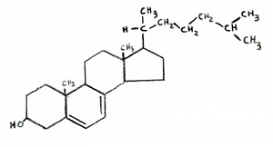 Vitamin D is synthesized from cholesterol in the skin, upon exposure to UV rays from the sun. In fact, the chemical formula of vitamin D is almost indistinguishable from that of cholesterol, as shown in the two attached figures (cholesterol on the left, vitamin D on the right). If LDL levels are
 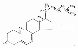 kept artificially low, then the body will be unable to resupply adequate amounts of cholesterol to replenish the stores in the skin once they have been depleted. This would lead to vitamin D deficiency, which is a widespread problem in America.
It is well known that vitamin D fights infection. To quote from [25], “Patients with severe infections as in sepsis have a high prevalence of vitamin D deficiency and high mortality rates.” As will be elaborated on later, a large number of infective agents have been shown to be present in abnormally high amounts in the brains of Alzheimers patients [27][26].
Dr. Grant has recently argued [16] that there are many lines of evidence pointing to the idea that dementia is associated with vitamin D deficiency. An indirect argument is that vitamin D deficiency is associated with many conditions that in turn carry increased risk for dementia, such as diabetes, depression, osteoporosis, and cardiovascular disease. Vitamin D receptors are widespread in the brain, and it is likely that they play a role there in fighting off infection. Vitamin D surely plays other vital roles in the brain as well, as powerfully suggested by this quote taken from the abstract of [32]: “We conclude there is ample biological evidence to suggest an important role for vitamin D in brain development and function.”
6. Astrocytes, Glucose Metabolism, and Oxygen
Alzheimer’s is clearly correlated with a deficiency in the supply of fat and cholesterol to the brain. IDL, when functioning properly, is actually incredibly efficient in cholesterol and fat throughput from the blood across cell membranes, compared to LDL [8]. It gives up its contents much more readily than the other apo’s. And it achieves this as a direct consequence of apoE. IDL (as well as LDL) in the blood delivers fats and cholesterol to the astrocytes in the brain, and the astrocytes can thus use this external source instead of having to produce these nutrients themselves. I suspect, in fact, that astrocytes only produce a private supply when the external supply is insufficient, and they do so reluctantly.
Why would it be disadvantageous for an astrocyte to synthesize its own fats and cholesterol? In my opinion, the answer has to do with oxygen. An astrocyte needs a significant energy source to synthesize fats and cholesterol, and this energy is usually supplied by glucose from the blood stream. Furthermore, the end-product of glucose metabolism is acetyl-Coenzyme A, the precursor to both fatty acids and cholesterol. Glucose can be consumed very efficiently in the mitochondria, internal structures within the cell cytoplasm, via aerobic processes that require oxygen. The glucose is broken down to produce acetyl-Coenzyme A as an end-product, as well as ATP, the source of energy in all cells.
However, oxygen is toxic to lipids (fats), because it oxidizes them and makes them rancid. Lipids are fragile if not encased in a protective shell like IDL, HDL, or LDL. Once they are rancid they are susceptible to infection by invasive agents like bacteria and viruses. So an astrocyte trying to synthesize a lipid has to be very careful to keep oxygen out, yet oxygen is needed for efficient metabolism of glucose, which will provide both the fuel (ATP) and the raw materials (acetyl-Coenzyme A) for fat and cholesterol synthesis.
What to do? Well, it turns out that there is an alternative, although much less efficient, solution: to metabolize glucose anaerobically directly in the cytoplasm. This process does not depend on oxygen (a great advantage) but it also yields substantially less ATP (only 6 ATP as contrasted with 30 if glucose is metabolized aerobically in the mitochondria). The end product of this anaerobic step is a substance called pyruvate, which could be further broken down to yield a lot more energy, but this process is not accessible to all cells, and it turns out that the astrocytes need help for this to happen, which is where amyloid-beta comes in.
7. The Crucial Role of Amyloid-Beta
Amyloid-beta (also known as “abeta”) is the substance that forms the famous plaque that accumulates in the brains of Alzheimer’s patients. It has been believed by many (but not all) in the research community that amyloid-beta is the principal cause of Alzheimer’s, and as a consequence, researchers are actively seeking drugs that might destroy it. However, amyloid-beta has the unique capability of stimulating the production of an enzyme, lactate dehydrogenase, which promotes the breakdown of pyruvate (the product of anaerobic glucose metabolism) into lactate, through an anaerobic fermentation process, with the further production of a substantial amount of ATP.
The lactate, in turn, can be utilized itself as an energy source by some cells, and it has been established that neurons are on the short list of cell types that can metabolize lactate. So I conjecture that the lactate is transported from the astrocyte to a neighboring neuron to enhance its energy supply, thus reducing its dependence on glucose. It is also known that apoE can signal the production of amyloid-beta, but only under certain poorly understood environmental conditions. I suggest those environmental triggers have to do with the internal manufacture of fats and cholesterol as opposed to the extraction of these nutrients from the blood supply. I.e., amyloid-beta is produced as a consequence of environmental oxidative stress due to an inadequate supply of fats and cholesterol from the blood.
In addition to being utilized as an energy source by being broken down to lactate, pyruvate can also be used as a basic building block for synthesizing fatty acids. So anaerobic glucose metabolism, which yields pyruvate, is a win-win-win situation: (1) it significantly reduces the risk of exposure of fatty acids to oxygen, (2) it provides a source of fuel for neighboring neurons in the form of lactate, and (3) it provides a basic building block for fatty acid synthesis. But it depends upon amyloid-beta to work.
Thus, in my view (and in the view of others [28] [20] Amyloid-Beta and Alzheimer’s), amyloid-beta is not a cause of Alzheimer’s, but rather a protective device against it. The abstract of reference [28] arguing this point of view is reproduced in full in the Appendix. Several variants of a genetic defect associated with amyloid precursor protein (APP), the protein from which amyloid-beta is derived, have now been identified. A defect in this protein, which is associated with an increased risk of early onset Alzheimer’s, would likely lead to a reduced ability to synthesize amyloid-beta, which would then leave the brain with a big problem, since both the fuel and the basic building blocks for fatty acid synthesis would be in short supply, while oxygen trekking through the cell to the mitochondria would be exposing whatever fats were being synthesized to oxidation. The cell would likely be unable to keep up with need, and this would lead to a reduction in the number of fatty acids in the Alzheimer’s cerebrospinal fluid, a well-established characteristic of Alzheimer’s [38].
8. Cholesterol’s Role in the Brain
The brain comprises only 2% of the body’s total weight, yet it contains nearly 25% of the total cholesterol in the body. It has been determined that the limiting factor allowing the growth of synapses is the availability of cholesterol, supplied by the astrocytes. Cholesterol plays an incredibly important role in the synapse, by shaping the two cell membranes into a snug fit so that the signal can easily jump across the synapse [50]. So inadequate cholesterol in the synapse will weaken the signal at the outset, and inadequate fat coating the myelin sheath will further weaken it and slow it down during transport. A neuron that can’t send its messages is a useless neuron, and it only makes sense to prune it away and scavenge its contents.
The neurons that are damaged in Alzheimer’s are located in specific regions of the brain associated with memory and high level planning. These neurons need to transmit signals long distances between the frontal and prefrontal cortex and the hippocampus, housed in the midbrain. The transport of these signals depends on a strong and tight connection in the synapse, where the signal is transferred from one neuron to another, and a secure transmission across the long nerve fiber, a part of the white matter. The myelin sheath which coats the nerve fiber consists mainly of fatty acids, along with a substantial concentration of cholesterol. If it is not well insulated, the signal transmission rate will slow down and the signal strength will be severely reduced. Cholesterol is crucial for the myelin as well as for the synapse, as demonstrated dramatically through experiments conducted on genetically defective mice by Gesine Saher et al. [45]. These mutant mice lacked the ability to synthesize cholesterol in myelin-forming oligodendrocytes. They had severly disturbed myelin in their brains, and exhibited ataxia (uncoordinated muscle movements) and tremor. In the abstract, the authors wrote unequivocally, “This shows that cholesterol is an indispensable component of myelin membranes.”
In a post-mortem study comparing Alzheimer’s patients with a control group without Alzheimer’s, it was found that the Alzheimer’s patients had significantly reduced amounts of cholesterol, phospholipids (e.g, B-HDL), and free fatty acids in the cerebrospinal fluid than did the controls [38]. This was true irrespective of whether the Alzheimer’s patients were typed as apoE-4. In other words, reductions in these critical nutrients in the spinal fluid are associated with Alzheimer’s regardless of whether the reduction is due to defective apoE. The reductions in fatty acids were alarming: 4.5 micromol/L in the Alzheimer’s patients, compared with 28.0 micromol/L in the control group. This is a reduction by more than a factor of 6 in the amount of fatty acid available to repair the myelin sheath!
People with the apoE-4 allele tend to have high serum cholesterol. The question of whether this high cholesterol level might be an attempt on the part of the body to adjust for a poor rate of cholesterol uptake in the brain was addressed by a team of researchers in 1998 [39]. They studied 444 men between 70 and 89 years old at the time, for whom there existed extensive records of cholesterol levels dating back to several decades ago. Most significantly, cholesterol levels fell for the men who developed Alzheimer’s prior to their showing Alzheimer’s symptoms. The authors suggested that their high cholesterol might have been a protective mechanism against Alzheimer’s.
One might wonder why their cholesterol levels fell. There was no mention of statin drugs in the article, but statins would certainly be an effective way to reduce cholesterol levels. The statin industry would like people to believe that high cholesterol is a risk factor for Alzheimer’s, and they are quite thrilled that high cholesterol early in life is correlated with Alzheimer’s much later. But these results suggest quite the opposite: that blood cholesterol levels are kept high intentionally by the body regulatory mechanisms in an attempt to compensate for the defect. A high concentration will lead to an increase in the rate of delivery to the brain, where it is critically needed to keep the myelin sheath healthy and to promote neuron signaling in the synapses.
Using MRI technology, researchers at UCLA were able to measure the degree of breakdown of myelin in specific regions of the brain [6]. They conducted their studies on over 100 people between 55 and 75 years old, for whom they also determined the associated apoE allele (2, 3, or 4). They found a consistent trend in that apoE-2 had the least amount of degradation, and apoE-4 had the most, in the frontal lobe region of the brain. All of the people in the study were thus far healthy with respect to Alzheimer’s. These results show that premature breakdown of myelin sheath (likely due to an insufficient supply of fats and cholesterol to repair it) is associated with apoE-4.
To summarize, I hypothesize that, for the apoE-4 Alzheimer’s patients, defective apoE has led to an impaired ability to transport fats and cholesterol from the blood stream, via the astrocytes, into the cerebrospinal fluid. The associated high blood serum cholesterol is an attempt to partially correct for this defect. For the rest of the Alzheimer’s patients (the ones without the apoE-4 allele but who also have severely depleted fatty acids in their cerebrospinal fluid), we have to look for another reason why their fatty acid supply chain might be broken.
9. Infections and Inflammation
To summarize what I have said so far, Alzheimer’s appears to be a consequence of an inability of neurons to function properly, due to a deficiency in fats and cholesterol. A compounding problem is that the fats over time will become rancid if they cannot be adequately replenished. Rancid fats are vulnerable to attack by microorganisms such as bacteria and viruses. Amyloid-beta is part of the solution because it allows the astrocytes to be much more effective in utilizing glucose anaerobically, which protects the internally synthesized fats and cholesterol from toxic oxygen exposure, while at the same time providing the energy needed both by the astrocyte for the synthesis process and by neighboring neurons to fuel their signal firings.
Besides the astrocytes, the microglia in the brain are also implicated in Alzheimer’s. Microglia promote neuron growth when all is well, but trigger neuron programmed cell death in the presence of toxic substances secreted by bacteria such as polysaccharides [56]. Microglia will defensively secrete cytokines (communication signals that promote an immune response) when exposed to infective agents, and these in turn will lead to inflammation, another well-known feature associated with Alzheimer’s [1]. The microglia are able to control whether neurons should live or die, and they surely base this decision on factors related to how well the neuron functions and whether it is infected. Once enough neurons have been programmed for cell death, the disease will manifest itself as cognitive decline.
10. Evidence that Infection is Associated with Alzheimer’s
There is substantial evidence that Alzheimer’s is related to an increased likelihood of infective agents appearing in the brain. Some researchers believe that infective agents are the principle cause of Alzheimer’s. There are a number of bacteria that reside in the human digestive system and can co-exist with our own cells without any harm. However, H. pylori, one that is quite common, has been recently shown to be responsible for stomach ulcers. It has been suspected that H. Pylori might be implicated in Alzheimer’s, and, indeed, a recent study showed that Alzheimer’s patients had a significantly higher concentration of an antibody against H. Pylori in both their cerebrospinal fluid and their blood than non-Alzheimer’s controls [26]. H. pylori was detected in 88% of the Alzheimer’s patients but only 47% of the controls. In an effort to treat the Alzheimer’s patients, the researchers administered a potent combination of antibiotics, and assessed the degree of mental decline over the next two years [27]. For 85% of the patients, the infection was successfully routed, and for those patients, cognitive improvement was also detected after two years had elapsed. So this was a nice example of the possibility of treating Alzheimer’s through antibiotics.
C. pneumoniae is a very common bacterium, estimated to infect 40-70% of adults. But there’s a big difference between a bacterium being in the blood stream and making its way into the inner sanctum of the brain. A study of post-mortem samples from various regions of the brains of Alzheimer’s patients and non-Alzheimer’s controls revealed a remarkably different statistic: 17 out of 19 Alzheimer’s brains tested positive for the bacterium, whereas only 1 out of 19 brains from the control group tested positive [5].
Many other infective agents, both viruses and bacteria, have been found to be associated with Alzheimer’s, including herpes simplex virus, picornavirus, Borna disease virus, and spirochete [23]. One proposal was that a particularbacteriophage — a virus that infects the bacterium C. pneumoniae — might be responsible for Alzheimer’s [14]. The authors argued that the phages might make their way into the mitochondria of the host cell and subsequently initiate Alzheimer’s.
11. Ketogenic Diet as Treatment for Alzheimer’s
One of the promising new treatment paradigms for Alzheimer’s is to have the patient switch to an extremely high fat, low carb diet, a so-called “ketogenic” diet. The name comes from the fact that the metabolism of dietary fats produces “ketone bodies” as a by-product, which are a very useful resource for metabolism in the brain. It is becoming increasingly clear that defective glucose metabolism in the brain (so-called “type-3 diabetes”) is an early characteristic of Alzheimer’s. Ketone bodies, whether they enter the astrocyte directly or are produced in the astrocyte itself by breaking down fats, can be delivered to adjacent neurons, as shown in the accompanying figure.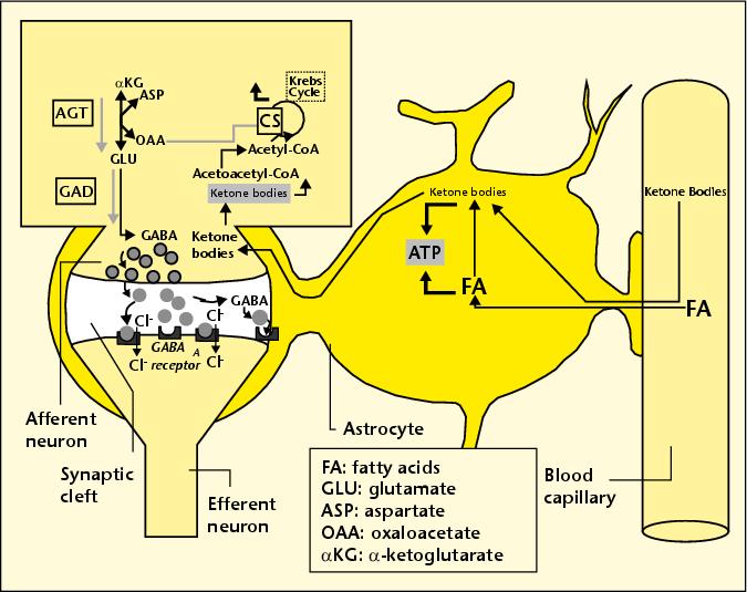 These neurons can utilize the ketone bodies both as an energy source (replacing and therefore relieving glucose) and as a precursor to GABA, a critical neurotransmitter that is widespread in the brain.
Evidence that a ketogenic diet might help Alzheimer’s was first found through research conducted on mice who had been bred to be prone to Alzheimer’s disease [21]. Researchers found that the mice’s cognition improved when they were treated with a high-fat low-carb diet, and also that the amount of amyloid-beta in their brain was reduced. The latter effect would be anticipated based on the premise that amyloid-beta promotes full utilization of glucose anaerobically, as I discussed previously. By having ketone bodies as an additional source of fuel, the dependence on glucose is reduced. But another effect that may be more important than this is the availability of high-quality fats to improve the condition of the myelin sheath.
This idea is supported by other experiments done on human Alzheimer’s patients [11] [42]. A placebo-controlled 2004 study [42] of the effect of dietary fat enrichment on Alzheimer’s is especially informative, because it uncovered a significant difference in effectiveness for the fat-enrichment for subjects who did not have the apoE-4 allele as compared with those who did. The experimental test group were given a supplemental drink containing emulsified medium chain triglycerides, found in high concentration in coconut oil. The subjects without the apoE-4 allele showed a significant improvement in score on a standard test for Alzheimer’s, whereas those with the apoE-4 allele did not. This is a strong indicator that the benefit may have to do with an increase in uptake by the astrocyte of these high-quality fats, something that the subjects with the apoE-4 allele are unable to accomplish due to the defective IDL and LDL transport mechanisms.
12. NADH Treatment: the Crucial Role of Antioxidants
One of the very few promising treatments for Alzheimer’s is the coenzyme, NADH (nicotinamide adenine dinucleotide) [12]. In a placebo-controlled study, Alzheimer’s subjects given NADH for six months exhibited significantly better performances on verbal fluency, visual constructional ability and abstract verbal reasoning than the control subjects given a placebo.
Why would NADH be effective? In the process of converting pyruvate to lactate, lactate dehydrogenase consumes oxygen by oxidizing NADH to NAD+, as illustrated in the accompanying figure. So, if the bioavailability of NADH is increased, it stands to reason that the astrocyte would have an enhanced ability to convert pyruvate to lactate, the critical step in the anaerobic metabolic pathway that is enhanced by amyloid-beta. The process, by absorbing the toxic oxygen, would reduce the damage to the lipids due to oxygen exposure, and would also provide lactate as a source of energy for the neurons.
13. Excessive Oxygen Exposure and Cognitive Decline
It has been observed that some elderly people suffer temporary and sometimes permanent cognitive decline following a lengthy operation. Researchers at the University of South Florida and Vanderbilt University suspected that this might be due to excessive exposure to oxygen [4]. Typically, during an operation, people are often administered high doses of oxygen, even as much as 100% oxygen. The researchers conducted an experiment on young adult mice, which had been engineered to be predisposed towards Alzheimer’s but had not yet suffered cognitive decline. They did however already have amyloid-beta deposits in their brains. The re-engineered mice, as well as a control group that did not have the Alzheimer’s susceptibility gene, were exposed to 100-percent oxygen for a period of three hours, three times over the course of several months, simulating repeated operations. They found that the Alzheimer’s pre-disposed mice suffered significant cognitive decline following the oxygen exposure, by contrast with the control mice.
This is a strong indication that the excessive oxygen exposure during operations is causing oxidative damage in the Alzheimer’s brain. Given the arguments I have presented above, this result makes good sense. The brain, by converting to anaerobic metabolism for generating energy (with help from amyloid-beta) is trying its best to avoid exposing the fatty acids and cholesterol to oxidative damage. But an extremely high concentration of oxygen in the blood makes it very difficult to protect the fats and cholesterol during transport through the blood, and also probably causes an unavoidable increase in oxygen uptake and therefore exposure within the brain itself.
14. Fats are a Healthy Choice!
You would practically have to be as isolated as an Australian Aborigine not to have absorbed the message that dietary fats, particularly saturated fats, are unhealthy. I am extremely confident that this message is false, but it is nearly impossible to turn the opinion tide due to its pervasive presence. Most people don’t question why fats are bad; they assume that researchers must have done their homework, and they trust the result.
To say that the current situation with regard to dietary fats is confusing would be an understatement. We are repeatedly told to keep our total fat intake down to, ideally, 20% of our total calories. This is difficult to achieve, and I believe it is misguided advice. In direct contradiction to this “low-fat” goal, we are encouraged to consume as much as possible of the “good” kinds of fats. Fortunately, the message is finally becoming widely embraced that omega-3 fats are healthy and that trans fats are extremely unhealthy. DHA (docosahexaenoic acid) is an omega-3 fat that is found in large quantities in the healthy brain. In the diet, it is available mainly from cold water fish, but eggs and dairy are also good sources. Trans fats are generated by a high-heat process that hydrolyzes polyunsaturated fats into a more stable configuration, which increases their shelf life but makes them so unnatural they almost can no longer be called a food. Trans fats are extremely damaging both to heart and brain health. A high consumption of trans fats has recently been shown to increase the risk of Alzheimer’s [41]. Trans fats are especially prevalent in highly processed foods — particularly when fats are converted to a powdered form.
We are told to avoid saturated fats, mainly because they have appeared, from empirical evidence, to be more likely to raise LDL levels than unsaturated fats. Yet these fats are less susceptible to oxidation, and this may be why they show up in LDL — because they are of higher quality and therefore should preferentially be delivered to the tissues for functional roles rather than as fuel (i.e., free fatty acids). Coconut oil, a saturated fat, has been shown to benefit Alzheimer’s patients [42]. And high-fat dairy (also highly saturated) has been shown to be beneficial both to fertility among women [10] and, remarkably, to heart disease [37][22].
Despite the widespread belief that fats (particularly saturated fats) are unhealthy, an article that appeared in the American Journal of Clinical Nutrition in 2004 [37] claims that, for a group of post-menopausal women, a high-fat, high-saturated-fat diet affords better protection from coronary artery disease than a low-fat (25% of calories from fats) diet. The subjects in the study were obese women with coronary artery disease. Most of them had high blood pressure, and many had diabetes. They fit the profile for metabolic syndrome that I have previously argued is a direct consequence of a prolonged low-fat high-carb diet. I am gratified to see that my hypothesis that an increase in fat intake would decrease their risk of heart disease has been verified by a carefully controlled study.
Another investigation where fats were shown to afford protection against heart disease has just been completed. It involved a long-term study of a large number of Swedish men [22]. The authors looked at low- vs high-fat dairy, as well as consumption of fruits and vegetables, meats, grains, etc. The only statistically significant result that afforded protection from heart disease was a combination of high-fat dairy and lots of fruits and vegetables. Fruits and vegetables with low-fat dairy afforded no protection.
I suspect one of the critical nutrients the fruits and vegetables provide is antioxidants that help prolong the life of the fats. Other excellent sources of antioxidants include richly colored fruits like berries and tomatoes, coffee, green tea, and dark chocolate, and several spices, most especially cinnamon and turmeric (a major ingredient of curry). These should be consumed in abundance along with fats for optimal results.
Polyunsaturated fats such as corn oil and canola oil are unhealthy for the brain precisely because they are unsaturated. There are two major problems: (1) they have a low melting point, which means that, if they are used for frying they will be converted to trans fats, which are extremely unhealthy, and (2) they are much more susceptible to becoming rancid (oxidized) at room temperature than saturated fats, i.e., they have a shorter shelf life.
Researchers in Germany recently conducted an ingenious experiment designed to determine how the degree of freshness of polyunsaturated fats affects the metabolism of those fats in female lactating rats [43]. They divided female rats into two groups, and the only difference between the test group and the controls was that the test group was given fats that had been left in a relatively warm place for 25 days, which caused considerable oxidative damage, whereas the controls were fed fresh fats instead. The rats’ unusual diet was begun on the day that they gave birth to a litter. The researchers examined the mammary glands and the milk produced by the two groups for apparent differences. They found that the test group’s milk was markedly reduced in the amount of fat it contained, and their mammary glands correspondingly took up less fat from the blood supply. One might surmise that the rats’ metabolic mechanisms were able to detect oxidative damage to the fats, and therefore rejected them, prefering to do without rather than to risk the consequences of feeding their pups oxidized fats. Consequently, the pups of the test group gained significantly less weight than the control group’s pups.
Boxed items like cookies and crackers that contain processed polyunsaturated fats are doctored with antioxidants and even antibiotics to protect them from spoiling. Once they’re consumed, however, they still have to be protected from going rancid. Biochemical laws work the same way whether inside or outside the body. There are plenty of bacteria throughout the body that would be eager to take up house-keeping in rancid fats. The body has devised all kinds of strategies for protecting fats from oxidation (becoming rancid) and from attack by bacteria. But its task is rendered much easier for saturated rather than unsaturated fats, and for fresh rather than stale fats.
If we stop trying to get by on as few fats as possible in the diet, then we don’t have to become so preoccupied with getting the “right” kinds of fats. If the body is supplied with an overabundance of fats, it can pick and choose to find the perfect fat to match each particular need; excess or defective fats can just be used as fuel, where it’s not very important which fat it is, as long as it can be broken down to release energy.
15. Summary and Conclusion
This is an exciting time for Alzheimer’s research, as new and surprising discoveries are coming out at a rapid pace, and evidence is mounting to support the notion that Alzheimer’s is a nutritional deficiency disease. It is an indication of how much progress has been made in recent years to note that 42% of the references in this essay were published in 2008 or 2009. A popular new theory is that Alzheimer’s may grow out of an impaired ability to metabolize glucose in the brain. The term “type-3 diabetes” has been coined to describe this defect, which often appears long before any symptoms of Alzheimer’s [49]. A shift from aerobic towards anaerobic glucose metabolism in the brain seems to be a harbinger of Alzheimer’s later in life, but I argue that the reason for this shift is both to provide a basic ingredient (pyruvate) from which to synthesize fatty acids, while simultaneously protecting them from potentially damaging oxidation. The ApoE-4 allele, which is associated with increased risk to Alzheimer’s, clearly implicates defects in fat and cholesterol transport, and the remarkable 6-fold reduction in the amount of fatty acids present in the cerebrospinal fluid of Alzheimer’s patients [38] speaks loudly the message that fat insufficiency is a key part of the picture. The observation that the myelin is degraded in the frontal lobes of the brains of people possessing the apoE-4 allele further substantiates the theory that the myelin repair mechanism is defective.
Cholesterol obviously plays a vital role in brain function. A whopping 25% of the total cholesterol in the body is found in the brain, and it is present in abundance both in the synapses and in the myelin sheath. The cholesterol in both of these places has been shown to play an absolutely essential role in signal transport and in growth and repair.
Given the strong positive role played by cholesterol, it can only be assumed that statin drugs would increase the risk of developing Alzheimer’s. However, the statin industry has been remarkably successful thus far in hiding this painful fact. They have managed to make much of the observation that high cholesterol much earlier in life is associated with an increased risk to Alzheimer’s thirty years later. Yet they offer not a single study, not even a retrospective study, to substantiate any claim that actively reducing cholesterol through statin therapy would improve the situation for these people. In fact, most damningly, the statin usage evidence that would answer the question was “unavailable” to the researchers who conducted the seminal study.
Beatrice Golomb is an M.D. Ph.D. who heads up the UCSD Statin Study group, a research team who are actively investigating the risk-benefit balance of statin drugs. She is increasingly becoming convinced that statin drugs should not be recommended for the elderly: that in their case the risks clearly outweigh the benefits. She makes a strong case for this position in an on-line article available here [15]. The section on Alzheimer’s is particularly compelling, and it points out the pitfalls in relying on previous studies done by the statin industry, where often those who have memory problems as side-effects of the statin drugs are excluded from the study, so that the results end up inappropriately biased in favor of statins. In summary, she wrote: “It must be emphasized that the randomized trial evidence has, to date, uniformly failed to show cognitive benefits by statins and has supported no effect or frank and significant harm to cognitive function.”
In addition to refusing to take statin therapy, another way in which an individual can improve their odds against Alzheimer’s is to consume plenty of dietary fats. It seems odd to suddenly switch from a “healthy” low-fat diet to an extremely high fat ketogenic diet, once a diagnosis of Alzheimer’s is made. A ketogenic diet consists, ideally, of 88% fat, 10% protein, and 2% carbohydrate [11]. That is to say, it is absurdly high in fat content. It seems much more reasonable to aim for something like 50% fat, 30% protein, and 20% carbohydrate, so as to pro-actively defend against Alzheimer’s.
I highly recommend a recent book written by the pediatric brain surgeon, Larry McCleary, M.D., called The Brain Trust Program [33]. This book gives a wealth of fascinating information about the brain, as well as specific recommendations for ways to improve cognitive function and avert later Alzheimer’s. Most significantly, he recommends a diet that is high in cholesterol and animal fats, including an abundance of fish, seafood, meat, and eggs. He also recommends coconuts, almonds, avocados and cheese, all foods that contain a significant amount of fat, while encouraging the avoidance of “empty carbs.” His knowledge on this subject grew out of his interest in helping his young patients heal more rapidly after brain trauma.
Our nation is currently bracing itself for an onslaught of Alzheimer’s, at a time when baby boomers are approaching retirement, and our health care system is already in a crisis of escalating costs and shrinking funds. We can not afford the high cost of caring for the swelling population of Alzheimer’s patients that our current practices of low-fat diet and ever expanding statin usage are promoting.
Appendix In this appendix, I include the full abstract of two papers that are relevant to the theory presented here. The first is the abstract of reference [19] in [46], which is reference [44] here [see the section on statin drugs above for context]:
Abstract, “Epidemiological and clinical trials evidence about a preventive role for statins in Alzheimer’s disease:”
“This paper reviews epidemiological and clinical trials data about whether statin use reduces the risk of Alzheimer’s disease (AD). The available information has come in three waves. The initial, mostly cross-sectional observational reports suggested that statins might prevent dementia. Next, two large clinical trials with cognitive add-on studies showed no benefit and neither did the third wave, again with observational studies. The latter were mostly longitudinal, and were critical of the first studies for not adequately addressing confounding by indication (i.e. that patients with dementia would be denied statins). Most recently, new data from the Canadian Study of Health and Aging have produced a mixed result. While methodological considerations are clearly important in understanding why the reports are so variable, there might also be merit in differentiating between statins, based on their presumed – and variable – mechanisms of action in dementia prevention, before concluding that the initial reports are entirely artefactual. Still, the first reports appear to have overestimated the extent of protection, so that unless there are important effects achievable with specific statins, a more than a modest role for statins in preventing AD seems unlikely.” The second abstract is taken from reference [28], on the “alternative hypothesis” that amyloid-beta is protective rather than detrimental to Alzheimer’s, i.e., that it is a “protective response to neuronal insult:”
Abstract, “Amyloid-beta in Alzheimer disease: the null versus the alternate hypotheses:”
“For nearly 20 years, the primary focus for researchers studying Alzheimer disease has been centered on amyloid-beta, such that the amyloid cascade hypothesis has become the “null hypothesis.” Indeed, amyloid-beta is, by the current definition of the disease, an obligate player in pathophysiology, is toxic to neurons in vitro, and, perhaps most compelling, is increased by all of the human genetic influences on the disease. Therefore, targeting amyloid-beta is the focus of considerable basic and therapeutic interest. However, an increasingly vocal group of investigators are arriving at an “alternate hypothesis” stating that amyloid-beta, while certainly involved in the disease, is not an initiating event but rather is secondary to other pathogenic events. Furthermore and perhaps most contrary to current thinking, the alternate hypothesis proposes that the role of amyloid-beta is not as a harbinger of death but rather a protective response to neuronal insult. To determine which hypothesis relates best to Alzheimer disease requires a broader view of disease pathogenesis and is discussed herein.”
Contact: seneff@csail.mit.edu
References
[1] H. Akiyama, S. Barger, S. Barnum, B. Bradt, J.Bauer, G.M. Cole, N.R. Cooper, P. Eikelenboom, M. Emmerling, B.L. Fiebich, C.E. Finch, S. Frautschy, W.S. Griffin, H. Hampel, M. Hull, G. Landreth, L. Lue, R. Mrak, I.R. Mackenzie, P.L. McGeer, M.K. O’Banion, J. Pachter, G. Pasinetti, C. Plata-Salaman, J. Rogers, R.Rydel, Y. Shen, W. Streit, R. Strohmeyer, I. Tooyoma, F.L. Van Muiswinkel, R. Veerhuis, D. Walker, S. Webster, B. Wegrzyniak, G. Wenk, and T. Wyss-Coray, “Inflammation and Alzheimer’s disease.” Neurobiol Aging (2000) May-Jun;21(3):383-421,
[2] Alzheimer’s Association, “Alzheimer’s Disease Facts and Figures,” Alzheimer’s and Dementia (2009) Vol. 5, Issue 3.
[3] K.J. Anstey, D.M. Lipnicki and L.F. Low, “Cholesterol as a risk factor for dementia and cognitive decline: a systematic review of prospective studies with meta-analysis.” Am J Geriatr Psychiatry (2008) May, Vol. 16, No. 5, pp. 343-54.
[4] G. Arendash, A. Cox, T. Mori, J. Cracchiolo, K. Hensley, J. Roberts 2nd, “Oxygen treatment triggers cognitive impairment in Alzheimer’s transgenic mice,” Neuroreport. (2009) Jun 18.
[5] B.J. Balin, C.S. Little, C.J. Hammond, D.M. Appelt, J.A. Whittum-Hudson, H.C. Gerard, A.P. Hudson, “Chlamydophila pneumoniae and the etiology of late-onset Alzheimer’s disease.” J. Alz. Dis. (2008) Vol. 13, pp. 371-380.
[6] G. Bartzokis, MD; P.H. Lu, Psy, D.H. Geschwind, MD, N.Edwards, MA, J. Mintz, PhD, and J.L. Cummings, MD, “Apolipoprotein E Genotype and Age-Related Myelin Breakdown in Healthy Individuals: Implications for Cognitive Decline and Dementia,” Arch Gen Psychiatry (2006) Vol. 63, pp. 63-72.
[7] N. Bernoud, L. Fenart, C. Bénistant, J. F. Pageaux, M. P. Dehouck, P. Molière, M. Lagarde, R. Cecchelli,d, and J. Lecerf, “Astrocytes are mainly responsible for the polyunsaturated fatty acid enrichment in blood-brain barrier endothelial cells in vitro” Journal of Lipid Research (1998) Sept., Vol. 39, pp. 1816-1824.
[8] M. S. Brown and J. L. Goldstein, “A Receptor-Mediated Pathway for Cholesterol Homeostasis,” Nobel Lecture, December 9, 1985.
[9] N. Cartier, C. Sevin, A. Benraiss, P. DeDeyn, D. Bonnin, M-T Vanier, M. Philippe, V. Gieselmann and P. Aubourg, “AAV5-Mediated Delivery of Human Aryl Sulfatase A (hARSA) Prevents Sufatide Storage and Neuropathological Phenotype in Metachromatic Leukodystrophy (MLD) Mice,” Molecular Therapy (2005) 11, S166-S167; doi: 10.1016/j.ymthe.2005.06.431
[10] J. Chavarro, W.C. Willett, and P.J. Skerrett, The Fertility Diet, (2008) McGraw Hill.
[11] L.C. Costantini, L.J. Barr, J.L. Vogel and S.T. Henderson, “Hypometabolism as a therapeutic target in Alzheimer’s disease” BMC Neurosci (2008) Vol. 9, Suppl. 2, S16. doi: 10.1186/1471-2202-9-S2-S16.
[12] V. Demarin, S.S. Podobnik, D. Storga-Tomic and G. Kay, “Treatment of Alzheimer’s disease with stabilized oral nicotinamide adenine dinucleotide: A randomized, double-blind study” Drugs Exp Clin Res. (2004) Vol. 30, No. 1, pp. 27-33.
[13] R.B. DeMattos, R.P. Brendza, J.E. Heuser, M.Kierson, J.R. Cirrito, J. Fryer, P.M. Sullivan, A.M. Fagan, X. Han and D.M. Holtzman, “Purification and characterization of astrocyte-secreted apolipoprotein E and J-containing lipoproteins from wild-type and human apoE transgenic mice,” Neurochem Int. (2001) Nov-Dec;39(5-6):415-25. doi:10.1016/S0197-0186(01)00049-3.
[14] M. Dezfulian, M.A. ShokrgozarA, S. Sardari, K. Parivar and G. Javadi, “Can phages cause Alzheimer’s disease?” Med Hypotheses (2008) Nov;71(5):651-6.
[15] B.A. Golomb, M.D., Ph.D., “Statin Adverse Effects: Implications for the Elderly,” Geriatric Times (2004) May/June, Vol. V, Issue 3
[16] W.R. Grant, Ph.D., “Does Vitamin D Reduce the Risk of Dementia?” Journal of Alzheimer’s Disease (2009) May, Vol. 17, No. 1., pp. 151-9.
[17] Dr. Duane Graveline, Lipitor: Thief of Memory, Statin Drugs and the Misguided War on Cholesterol, (2004) http://www.buybooksontheweb.com.
[18] X. Han, “Potential mechanisms contributing to sulfatide depletion at the earliest clinically recognizable stage of Alzheimer’s disease: a tale of shotgun lipidomics,” J Neurochem (2007) November, Vol. 103, Suppl. 1. pp. 171-179. doi: 10.1111/j.1471-4159.2007.04708.x.
[19] X. Han, H. Cheng, J.D. Fryer, A.M. Fagan and D.M. Holtzman, “Novel Role for Apolipoprotein E in the Central Nervous System: Modulation of Sulfatide Content” Journal of Biological Chemistry, March 7, 2003, Vol. 278, pp. 8043-8051, DOI 10.1074/jbc.M212340200.
[20] K. Heininger, “A unifying hypothesis of Alzheimer’s disease. IV. Causation and sequence of events,” Rev Neurosci. (2000) Vol. 11, Spec No, pp.213-328.
[21] S.T. Henderson, “Ketone Bodies as a Therapeutic for Alzheimer’s Disease,” NeuroTherapeutics,, (2008) Jul;5(3):470-80, doi:10.1016/j.nurt.2008.05.004
[22] S. Holmberg, A. Thelin and E.-L. StiernstrNvm, “Food Choices and Coronary Heart Disease: A Population Based Cohort Study of Rural Swedish Men with 12 Years of Follow-up,” Int. J. Environ. Res. Public Health (2009) Vol. 6, pp. 2626-2638;
[23] K. Honjo, R. van Reekum, and N.P. Verhoeff, “Alzheimer’s disease and infection: do infectious agents contribute to progression of Alzheimer’s disease?” Alzheimers Dement. (2009) Jul;5(4):348-60.
[24] S.M. Innis and R.A. Dyer, “Brain astrocyte synthesis of docosahexaenoic acid from n-3 fatty acids is limited at the elongation of docosapentaenoic acid,” (2002) Sept. Journal of Lipid Research, Vol. 43, pp. 1529-1536.
[25] L. Jeng, A.V. Yamshchikov, S.E. Judd, H.M. Blumberg, G.S. Martin, T.R. Ziegler and V. Tangpricha, “Alterations in Vitamin D Status and Anti-microbial Peptide Levels in Patients in the Intensive Care Unit with Sepsis,” Journal of translational Medicine,” (2009) Vol. 7, No. 28.
[26] J. Kountouras, M. Boziki, E. Gavalas, C. Zavos, G. Deretzi, N. Grigoriadis, M. Tsolaki, D. Chatzopoulos, P. Katsinelos, D. Tzilves, A. Zabouri, I. Michailidou, “Increased cerebrospinal fluid Helicobacter pylori antibody in Alzheimer’s disease,” Int J Neurosci. (2009) 119(6):765-77.
[27] J. Kountouras, M. Boziki, E. Gavalas, C. Zavos, N. Grigoriadis, G. Deretzi, D. Tzilves, P. Katsinelos, M. Tsolaki, D. Chatzopoulos, and I. Venizelos, “Eradication of Helicobacter pylori may be beneficial in the management of Alzheimer’s disease,” J Neurol. (2009) May;256(5):758-67. Epub 2009 Feb 25.
[28] H.G. Lee, X. Zhu, R.J. Castellani, A. Nunomura, G. Perry, and M.A. Smith, “Amyloid-beta in Alzheimer disease: the null versus the alternate hypotheses,” J Pharmacol Exp Ther. (2007) June, Vol. 321 No. 3, pp. 823-9. doi:10.3390/ijerph6102626.
[29] J. Marcus, S. Honigbaum, S. Shroff, K. Honke, J. Rosenbluth and J.L. Dupree, “Sulfatide is essential for the maintenance of CNS myelin and axon structure,” Glia (2006), Vol. 53, pp. 372-381.
[30] R.T. Matthews, L. Yang, S. Browne, M. Baik and M.F. Beal, “Coenzyme Q10 administration increases brain mitochondrial concentrations and exerts neuroprotective effects,” Proc Natl Acad Sci U S A. (1998) Jul 21, Vol. 95, No. 15, pp.8892-7.
[31] D. Lutjohann and K. von Bergmann, “24S-hydroxycholesterol: a marker of brain cholesterol metabolism”Pharmacopsychiatry (2003) January 10, Vol. 36 Suppl 2, pp. S102-6, DOI: 10.1055/s-2003-43053.
[32] J. C. McCann and B.N. Ames, “Is there convincing biological or behavioral evidence linking vitamin D deficiency to brain dysfunction?”, (2008) FASEB J. Vol. 22, pp. 982-1001. doi: 10.1096/fj.07-9326rev.
[33] Larry McCleary, M.D., The Brain Trust Program (2007) September, The Penguin Group, New York, New York.
[34] B. McGuinness et al., “Statins for the prevention of dementia,” Cochrane Database of Systematic Reviews,(2009) No. 2.
[35] M.M. Mielke, P.P. Zandi, M. Sjogren, et al. “High total cholesterol levels in late life associated with a reduced risk of dementia,” Neurology (2005) Vol. 64, pp. 1689-1695.
[36] S.A. Moore, “Polyunsaturated Fatty Acid Synthesis and Release by Brain-Derived Cells in Vitro,” Journal of Molecular Neuroscience (2001), Vol. 16, pp. 195ff.
[37] D. Mozaffarian, E.B. Rimm, D.M. Herrington, “Dietary fats, carbohydrate, and progression of coronary atherosclerosis in postmenopausal women,” Am J Clin Nutr (2004) Vol. 80, pp. 1175-84.
[38] M. Mulder, R. Ravid, D.F. Swaab, E.R. de Kloet, E.D. Haasdijk, J. Julk, J.J. van der Boom and L.M. Havekes, “Reduced levels of cholesterol, phospholipids, and fatty acids in cerebrospinal fluid of Alzheimer disease patients are not related to apolipoprotein E4,” Alzheimer Dis Assoc Disord. (1998) Sep, Vol. 12, No. 3, pp. 198-203.
[39] I.L. Notkola, R. Sulkava, J. Pekkanen, T. Erkinjuntti, C. Ehnholm, P. Kivinen, J. Tuomilehto, and A. Nissinen, “Serum total cholesterol, apolipoprotein E epsilon 4 allele, and Alzheimer’s disease,” Neuroepidemiology (1998) Vol. 17, No. 1, pp. 14-20.
[40] F.W. Pfrieger, “Outsourcing in the brain: Do neurons depend on cholesterol delivery by astrocytes?”, BioEssays(2003) Vol. 25 Issue 1, pp.72-78.
[41] A. Phivilay, C. Julien, C. Tremblay, L. Berthiaume, P. Julien, Y. Giguère and F. Calon, “High dietary consumption of trans fatty acids decreases brain docosahexaenoic acid but does not alter amyloid-beta and tau pathologies in the 3xTg-AD model of Alzheimer’s disease.” Neuroscience (2009) Mar 3, Vol. 159, No. 1, pp. 296-307. Epub 2008 Dec 14.
[42] M.A. Reger, S. T. Henderson, C. Hale, B. Cholerton, L.D. Baker, G.S. Watson, K. Hyde, D. Chapman and S. Craft, “Effects of Beta-hydroxybutyrate on cognition in memory-impaired adults,” Neurobiology of Aging (2004) Vol. 25, No. 3, March, pp. 311-314,
[43] R. Ringseis, C. Dathe, A. Muschick, C. Brandsch and K. Eder, “Nutrient Physiology, Metabolism, and Nutrient-Nutrient Interactions Oxidized Fat Reduces Milk Triacylglycerol Concentrations by Inhibiting Gene Expression of Lipoprotein Lipase and Fatty Acid Transporters in the Mammary Gland of Rats,” American Society for Nutrition J. Nutr. (2007) Sept., Vol. 137, pp. 2056-2061.
[44] K. Rockwood, “Epidemiological and clinical trials evidence about a preventive role for statins in Alzheimer’s disease.” Acta Neurol Scand Suppl. (2006) Vol. 185, pp. 71-7.
[45] G. Saher, B. Brugger, C. Lappe-Siefke, W. Mobius, R. Tozawa, M.C. Wehr, F. Wieland, S. Ishibashi, and K.A. Nave, “High cholesterol level is essential for myelin membrane growth.” Nat Neurosci (2005) Apr, Vol. 8, No. 4, pp. 468-75. Epub 2005 Mar 27.
[46] A. Solomon, M. Kivipelto, B. Wolozin, J. Zhou, and R.A. Whitmer, “Midlife Serum Cholesterol and Increased Risk of Alzheimer’s and Vascular Dementia Three Decades Later,” Dementia and Geriatric Cognitive Disorders (2009) Vol. 28, pp. 75-80, DOI: 10:1159/000231980.
[47] M. Simons, MD, P. Keller, PhD, J. Dichgans, MD and J.B. Schulz, MD, “Cholesterol and Alzheimer’s disease: Is there a link?” Neurology (2001) Vol. 57, pp. 1089-1093.
[48] L.L. Smith, “Another cholesterol hypothesis: cholesterol as antioxidant,” Free Radic Biol Med. (1991) Vol. 11, No. 1, pp. 47-61.
[49] E. Steen, B.M. Terry, E.J. Rivera, J.L. Cannon, T.R. Neely, R. Tavares, X.J. Xu, J.R. Wands, and S.M. de la Monte “Impaired insulin and insulin-like growth factor expression and signaling mechanisms in Alzheimer’s disease – is this type 3 diabetes?” Journal of Alzheiner’s Disease (2005) Vol. 7, Number 1, pp. 63-80.
[50] J. Tong, P.P. Borbat, J.H. Freed and Y-K Shin, “A scissors mechanism for stimulation of SNARE-mediated lipid mixing by cholesterol,” PNAS (2009) March 31 Vol. 106, No. 13, pp. 5141-5146.
[51] M-C Vohl, T. A.-M. Neville, R. Kumarathasan, S. Braschi, and D.L. Sparks, “A Novel Lecithin-Cholesterol Acyltransferase Antioxidant Activity Prevents the Formation of Oxidized Lipids during Lipoprotein Oxidation,”Biochemistry (1999) Vol. 38 No. 19, pp. 5976-5981. DOI: 10.1021/bi982258w.
[52] M. Waldman, MD,, 9th International Conference on Alzheimer’s and Parkinson’s Diseases (2009) Abstract 90, Presented March 12-13.
[53] R. West, M.A., M. Schnaider Beeri, Ph.D., J. Schmeidler, Ph.D., C. M. Hannigan, B.S., G. Angelo, M.S., H.T. Grossman, M.D., C. Rosendorff, M.D., Ph.D., and J.M. Silverman, Ph.D., “Better memory functioning associated with higher total and LDL cholesterol levels in very elderly subjects without the APOE4 allele,” Am J Geriatr Psychiatry(2008) September; Vol. 16, No. 9, pp. 781-785. doi: 10.1097/JGP.0b013e3181812790.
[54] A.W.E. Weverling-Rijnsburger, G.J. Blauw, A.M. Lagaay, D.L. Knook, A.E. Meinders, and R.G.J. Westendorp, “Total cholesterol and risk of mortality in the oldest old,” The Lancet, (1997) Vol. 350, No. 9085, pp. 1119-1123,
[55] R.F. Wilson, J.F. Barletta and J.G. Tyburski, “Hypocholesterolemia in Sepsis and Critically Ill or Injured Patients”Critical Care (2003), Vol. 7, pp. 413-414.
[56] S.-C. Zhang and S. Fedoroff, “Neuron-microglia Interactions in Vitro,” Acta Neuropathol (1996) Vol. 91, pp. 385-395.
New Study Shows Vitamin D3 Can Inhibit COVID-19
By Dr. Joseph Mercola | September 19, 2021
A study using active forms of vitamin D3 has shown that the vitamin’s metabolites can inhibit replication and expansion of SARS-CoV-2, the virus that causes COVID-19.
Summarized by Newswise, “researchers on this study say their findings help explain a possible mechanism for why low vitamin D levels seem to promote COVID-19 infection and poor outcome in certain individuals. This correlates to other studies showing a relationship between vitamin D deficiency and poor disease outcomes. More studies and clinical trials are planned to test the efficacy of vitamin D and lumisterol as an antiviral therapeutic for COVID-19 in animals and humans.”
Study authors commented, “Active forms of vitamin D and lumisterol can inhibit SARS-CoV-2 replication machinery enzymes, which indicates that novel vitamin D and lumisterol metabolites are candidates for antiviral drug research.”
In September 2020, Dr. Anthony Fauci, director of the National Institute of Allergy and Infectious Diseases, said he believed that vitamin D could help fight COVID, although he didn’t elaborate at the time on how he knew that to be true.
SOURCES:
ROBERT MALONE INTERVIEWED BY JIMMY DORE
anti_republocrat | September 15, 2021
Robert Malone, inventor of mRNA technology, is interviewed by Jimmy Dore. Malone is not “anti-vax,” but he is “pro-ethics” and believes that all medical procedures require truly informed consent, with absolutely no coercion. He shares the view of Geert Vanden Bossche, whom he mentions in the interview, that the vaccines help to generate the variants because they are non-sterilizing. He says they should be targeted toward those who are at highest risk from the virus, seniors and those with multiple co-morbidities. I personally disagree with that. I think they should be taken off the market altogether, but at least he is adamantly against mandates.
The Conspiracy Theorists Were Right; It IS a “Poison-Death Shot”

BY MIKE WHITNEY • UNZ REVIEW • SEPTEMBER 16, 2021
“I’ll do one more mind experiment with you: If everyone on the planet were to get Covid and not get treated, the death-rate globally would be less than half a percent. I’m not advocating for that, because 35 million people would die. However, if we follow the advice of some of the global leaders– like Bill Gates who said last year said “7 billion people need to be vaccinated”– then the death-rate will be over 2 billion people! SO, WAKE UP! THIS IS WORLD WAR 3! We are seeing a level of malevolence that we haven’t seen in the history of humanity!” Dr. Vladimir Zelenko, Author of The Zelenko “Early Treatment” Protocol that saved thousands of Covid-19 patients. (“Zelenko schools the Rabbinic Court”, Rumble; start at 11:45 minutes)
Did the regulators at the FDA know that all previous coronavirus vaccines had failed in animal trials and that the vaccinated animals became either severely ill or died?
Yes, they did.
Did they know that previous coronavirus vaccines had a tendency to “enhance the infection” and “make the disease worse”?
Yes.
Did Dr Anthony Fauci know that coronavirus vaccines had repeatedly failed and increased the severity of the infection?
Yes, he did. (See here: Fauci on ADE)
Did the drug companies conduct any animal trials prior to the FDA’s approval that would have convinced a reasonable person that the vaccines were safe to use on humans?
No, they didn’t.
Did they complete long-term clinical trials to establish whether the vaccines were safe?
No, there were no long-term clinical trials.
Did they conduct any biodistribution studies that showed where the substance in the injection goes in the body?
They did, but the data was not made available to the public.
Do the contents of the vaccine largely collect in various organs and in the lining of the vascular system?
Yes, they do.
Do large amounts of the substance accumulate in the ovaries?
Yes.
Will this effect female fertility and a woman’s ability to safely bring a baby to term?
The drug companies are currently researching this. The results are unknown.
Does the vaccine enter the bloodstream and collect in the lining of the blood vessels forcing the cells to produce the spike protein?
Yes.
Is the spike protein a “biologically active” pathogen?
It is.
Does the spike protein cause blood clots and leaky blood vessels in a large percentage of the people that are vaccinated?
It does, although the blood clots are mostly microscopic and appear in the capillaries. Only a small percentage of vaccinees get strokes or suffer cardiac arrest.
Should people be made aware of these possible bad outcomes before they agree to get vaccinated? (“Informed consent”)
Yes.
Did the FDA know that Pfizer had “identified vaccine-associated enhanced disease, including vaccine-associated enhanced respiratory disease, as an important potential risk”?
Yes, they did, but they did not demand that Pfizer fix the problem. Here’s more:
“The FDA noted that Pfizer, “identified vaccine-associated enhanced disease, including vaccine-associated enhanced respiratory disease, as an important potential risk”. The EMA similarly acknowledged that “vaccine associated enhanced respiratory disease” was “an important potential risk… that may be specific to vaccination for COVID- 19”.
Why neither regulator sought to exclude such dangers prior to emergency use authorization is an open
question that all doctors and patients are entitled to ask. Why medical regulators failed to investigate the
finding that large vaccine particles cross blood vessel walls, entering the bloodstream and posing risks of blood clotting and leaky vessels is yet another open question again.” (“Open Letter to the EMA and European Parliament”, Doctors for Covid Ethics)
Did the drug companies vaccinate the people in the placebo group after the clinical trials in order to conceal the difference in the long-term health outcomes between the two groups?
That is the conclusion a rational person would make.
So, they nuked the trials?
Yes.
Did the FDA largely shrug-off its regulatory duties and abandon its normal standards and protocols because
a– It wanted to rush the Covid vaccines into service as rapidly as possible?
b– It knew the Covid-19 vaccine would never meet long-term safety standards?
We don’t know yet, but the adverse events report strongly suggests that the Covid-19 vaccine is hands-down the most dangerous vaccine in history.
Is the FDA rushing the “boosters” without proper testing?
Yes, it is. Here’s a clip from author Alex Berenson’s latest at Substack:
“Pfizer basically hasn’t bothered to test the booster AT ALL in the people actually at risk – it conducted a single “Phase 1” trial that covered 12 people over 65. The main Phase 2/3 booster trial (beware efforts to cover multiple “phases” of drug research at once, you want it bad you get it bad) included no one over 55.
No one.
As in NONE.” (“Are you kidding me, Pfizer, volume 1 gazillion”, Alex Berenson, Substack)
Have the boosters been modified or improved to meet the changes in Delta variant?
No.
Is there any additional risk in taking a booster-shot after already taking two experimental gene-based vaccines in less than a year?
Considerable risk. Here’s more from the Doctors for Covid Ethics:
“Given that booster shots repeatedly boost the immune response to the spike protein, they will progressively boost self-to-self immune attack, including boosting complement-mediated damage to vessel walls.
Clinically speaking, the greater the vessel leakage and clotting that subsequently occurs, the more likely that organs supplied by the affected blood flow will sustain damage. From stroke to heart attack to brain vein thrombosis, the symptoms can range from death to headaches, nausea and vomiting, all of which heavily populate adverse reactions to COVID-19 vaccines.
As well as damage from leakage and clotting alone, it is additionally possible that the vaccine itself may leak into surrounding organs and tissues. Should this take place, the cells of those organs will themselves begin to produce spike protein, and will come under attack in the same way as the vessel walls. Damage to major organs such as the lungs, ovaries, placenta and heart can be expected ensue, with increasing severity and frequency as booster shots are rolled out.” (“Open Letter to the EMA and European Parliament“, Doctors for Covid Ethics)
So, it’s the double-whammy. On the one hand, the booster will perform largely like the original vaccine, penetrating cells and forcing them to produce spike protein which, in turn, generates blood clots and leaky blood vessels. And, on the other, the newly-produced S proteins trigger a damaging immune response in which the complement system attacks and destroys the cells that line the inside of the blood vessels. Every additional booster will intensify this process weakening the vascular system and increasing the clotting. If the Doctors are correct in their analysis, then we could see a sharp uptick in all-cause mortality in the heavily-vaccinated countries in less than a year. Cardiac arrests are already rising.
Here’s another question that’s worth mulling over: Was there any reason for the regulators at the FDA to think that these problems would not arise following the launching of the vaccine campaign?
No. They should have known there would be problems as soon as they saw that the vaccine did not stay in the shoulder as it was supposed to. The vaccine wasn’t supposed to enter the bloodstream and spread across the body leaving billions of spike proteins in its wake. (The spike protein is a cytotoxin, a cell killer. It is not an appropriate antigen for stimulating an immune response. It is a potentially-lethal pathogen that poses a threat to one’s health even if it is separated from the virus.) Nor was the vaccine supposed to trigger Antibody-Dependent Enhancement (ADE) which is the condition we hinted at above when referring to “vaccine-associated enhanced disease”. Here’s a brief explanation:
“ADE has proven to be a serious challenge with coronavirus vaccines, and this is the primary reason many have failed in early in-vitro or animal trials. For example, rhesus macaques who were vaccinated with the Spike protein of the SARS-CoV virus demonstrated severe acute lung injury when challenged with SARS-CoV, while monkeys who were not vaccinated did not. Similarly, mice who were immunized with one of four different SARS-CoV vaccines showed histopathological changes in the lungs with eosinophil infiltration after being challenged with SARS-CoV virus. This did not occur in the controls that had not been vaccinated. A similar problem occurred in the development of a vaccine for FIPV, which is a feline coronavirus.” (“Is the Coronavirus Vaccine a Ticking-Time Bomb?”, Science with Dr. Doug)
Is this what we are seeing right now? In all the countries that launched mass-vaccination campaigns early (Israel, Iceland, Scotland, Gibraltar and UK) cases, hospitalizations and deaths are rising faster in the vaccinated portion of the population than the unvaccinated. Why?
Are they really experiencing a fourth or fifth wave or have the vaccines generated “inactivity-enhancing” antibodies that make the disease worse? This 2-minute video helps to clarify what’s going on:
“Vaccines are made to a specific variant. And when that variant mutates, the vaccine no longer recognizes it. It’s like you are seeing a completely new virus. And, because that is so, you actually get more severe symptoms when you are vaccinated against one variant and it mutates and then your body sees the other variant. The science shows, that if you get vaccinated in multiple years (for the flu), you are more likely to get severe disease, you are more likely to get viral replication, and you are more likely to be hospitalized… We are seeing the same thing in Covid with the Delta variant. So we are actually mandating that people get a vaccine when they can actually get more sick when they are exposed to the virus… In fact, this week, a paper came out that showed that–with the Delta variant– when you are vaccinated your body is supposed to make antibodies that neutralize the virus, but they were supposed to neutralize the old variant. When they see this new variant, the antibodies take the virus and help it infect the cells.” (“Expert testimony on mandatory vaccinations”, Dr Christina Parks PhD., Rumble, start at minute 5:05)
Repeat: “If you get vaccinated in multiple years, you are more likely to get severe disease, you are more likely to get viral replication, and you are more likely to be hospitalized…. With the Delta variant– when you are vaccinated …. the antibodies take the virus and help it infect the cells.”
This is ADE, and this is probably why hospitalizations and deaths are rising among the vaccinated in Israel, UK and the rest. True, the Delta variant is less lethal than the Wuhan virus but, unfortunately, that rule does not apply to those who have been vaccinated and whose antibodies promote the uptake of the virus into their cells. This increases the viral replication function that increases the severity of the disease. In short, people are getting sicker because they were vaccinated. Here’s another short video that helps to explain:
“… The vaccine-induced antibodies will stand up against the virus. and once a virus is under pressure; it changes, it becomes a variant, and the variant cannot be stopped by vaccine-induced antibodies. Vaccine-induced antibodies. also shut down your innate immune system… so variants can come straight through and infect those that are vaccinated. That is viral immune escape, and that means that the vaccinated are defenseless against variants. This is no longer a pandemic of Covid-19. It is a pandemic of variants…
And there is something called recombination, and recombination means a vaccinated host can be infected by more than one variant at a time. …If a vaccinated host is co-infected by more than one variant, the variants will mix DNA, and change and camouflage and produce a super variant. And if a super variants are produced, nothing can stop them. And already they are saying that the latest variant to come out is vaccine resistant. And this is just the beginning. Dr Geert Vanden Bosche warns that if we do not immediately stop mass vaccination campaigns around the world, the world will experience an international catastrophe of mass mortality. I didn’t say that, he did. The vaccinated are a threat to us all.” (“Viral Immune Escape Explained”, Dr. Michael McDowell, Rumble)
It’s not the variant that intensifies the disease, it’s the fact that the vaccine targets one narrow endpoint, the spike protein, that gradually adapts to survive. As the virus progressively learns to avoid the vaccine, vaccine-induced immunity wanes. Natural immunity produces broad, robust immunity to the whole virus not merely one part of it. It is strong and enduring.
So how will the vaccinated fight new forms of the virus, after all, the vaccine is not a medicine that overpowers a particular pathogen. It is a subtle (genetic) reprogramming of the immune system that forces one’s cells to produce a particular version of the spike protein. Boosters that stimulate production of the same protein will have only modest impact. In short, boosters are still fighting the last war.
Also, as we mentioned above, coronavirus vaccines tend to create antibodies that “enhance infectivity” when they encounter adapted forms of the virus. That means that millions of inoculated people will now face forms of the virus for which they have almost no protection and for which their compromised immune systems can only provide limited help. Here’s more from the article above:
“Right now, the fatality rate of the virus is estimated to be approximately 0.26%, and this number seems to be dropping as the virus is naturally attenuating itself through the population. It would be a great shame to vaccinate the entire population against a virus with this low of a fatality rate, especially considering the considerable risk presented by ADE. I believe this risk of developing ADE in a vaccinated individual will be much greater than 0.26%, and, therefore, the vaccine stands to make the problem worse, not better. It would be the biggest blunder of the century to see the fatality rate of this virus increase in the years to come because of our sloppy, haphazard, rushed efforts to develop a vaccine with such a low threshold of safety testing and the prospect of ADE lurking in the shadows.” (“Is the Coronavirus Vaccine a Ticking-Time Bomb?”, Science with Dr. Doug)
“Blunder”, he says?
It wasn’t a blunder. It was deliberate. The Covid-19 vaccine was supposed to fail like all the coronavirus vaccines before it. That’s the point. That’s why the drug companies skipped the animal testing and long-term safety trials. That’s why the FDA rushed it through the regulatory process and suppressed the other life-saving medications, and silenced all critics of the policy, and pushed for universal vaccination regardless of the risks of blood clotting, cardiac arrest, stroke and death. And that’s why the world is on the threshold of an “international catastrophe of mass mortality.” It’s because that’s how the strategy was planned from the very beginning.
The vaccine isn’t supposed to work, it’s supposed to make things worse. And it has! It’s increased the susceptibility of millions of people to severe illness and death. That’s what it’s done. It’s a stealth weapon in an entirely new kind of war; a war aimed at restructuring the global order and establishing absolute social control. Those are the real objectives. It has nothing to do pandemics or viral contagion. It’s about power and politics. That’s all.
Instagram blocks results for “natural immunity” hashtag
By Cindy Harper | Reclaim The Net | September 17, 2021
Instagram has blocked the results page for the use of the hashtag #naturalimmunity.
When the hashtag is selected, Instagram says, “This hashtag is hidden,” and that “Posts for #naturalimmunity have been limited because the community has reported some content that may not meet Instagram’s Community Guidelines.”
Many posts using the hashtag were centered around stories that suggest that those who have recovered from COVID were less likely to catch COVID again than someone who was vaccinated but had no prior exposure to COVID.
A 700,000-person Israeli study this month found those who had experienced prior infections were 27 times less likely to get a second symptomatic infection than those who were only vaccinated, and many have taken to social media to discuss it.
However, Instagram has started to censor the hashtag.
Congressman Thomas Massie, who has kept informed about Big Tech censorship, commented on the block, saying, “Instagram blocks #naturalimmunity hashtag. Don’t forget Congress gave @CDCgov $1 billion to market the vaccines. I suspect a lot of that has made its way into the hands of social media companies. Also, factcheck-dot-org is funded by a group that holds $2 billion of vaccine stock.”
Australia’s Labor Party asks Google what “misinformation” censorship plans it has for the next election

By Christina Maas | Reclaim The Net | September 17, 2021
Australia’s Labor Party wants Google to explain the steps it has taken to ensure its platforms are not “exploited for misinformation” ahead of the next general election. The party says it fears its rivals will use “misinformation” to gain an edge in the upcoming election.
According to The Guardian, Labor’s national secretary Paul Erickson sent a letter to Google Australia’s managing director Mel Silva, asking if the company has improved its systems since the last election in 2019 to “ensure its platforms and advertising capabilities are not exploited for misinformation.”
In the letter, Erickson mentions Craig Kelly and businessman Clive Palmer for their criticism of the strict COVID-19 measures. He notes several videos posted by Kelly on his YouTube account “in which Mr Kelly promotes ivermectin and hydroxychloroquine as effective treatments for COVID-19 or claims that Covid-19 vaccines are unsafe,” according to The Guardian.
Kelly, a former member of the Liberal Party, formed his own party, the United Australian Party (UAP).
Erickson’s letter further asks Google how it plans to handle “the elevated risk of misinformation in the context of the upcoming federal election, including in relation to content uploaded by the UAP.”
The Labor leader notes that the UAP “is already spending hundreds of thousands of dollars on political advertising, including on Google’s platforms.” He insisted that it was crucial for Google’s platforms not to be “misused” amid a pandemic, “including by those with a track record of spreading politically motivated misinformation in the lead-up to the next federal election.”
“Regrettably, the response of digital platforms was wholly inadequate,” Erickson wrote. “These mistakes should not be repeated.”
The Labor party was the victim of a misinformation campaign relating to the “death tax” in the last election.
Kelly slammed Erickson for the letter.
“It is a disgrace and a new low that a political party would ask a foreign oligarch to censor freedom of speech in Australian politics,” the MP told The Guardian Australia. “The idea that an alternate opinion of an expert is misinformation is a claim I categorically reject.”
The UAP leader described Erickson’s letter as “silencing of genuine debate, and that will leave the public misinformed.”
Why the Biden COVID-19 Vaccine Mandate is Unconstitutional
Techno Fog | September 14, 2021
On September 9, President Biden announced he would circumvent the democratic process, ordering the Secretary of the Department of Labor to require employers with over 100 workers to “ensure their workforces are fully vaccinated or show a negative test at least once a week.”
This was essential, as Biden said, “to protect vaccinated workers from unvaccinated workers.”
As we have explained, the Secretary of Labor will issue these regulations through OSHA by way of an Emergency Temporary Standard (ETS). The ETS would allow the Secretary of Labor to issue the vaccine mandate without the normal administrative rulemaking requirements (like notice and public comment periods).

While the Biden Administration tells the public that there’s no time to waste in issuing the mandate, the truth is that OSHA/Labor failed to argue the necessity of a vaccine mandate since the vaccines have been available – a time period approaching one year. Moreover, the Biden Department of Labor is secretly meeting with the US Chamber of Commerce and business lobbyists to gather support for the mandate. As Bloomberg Law reports:
Solicitor of Labor Seema Nanda held a virtual meeting with Neil Bradley, the Chamber’s chief policy officer, and other business lobbyists. The Chamber, the largest business lobbying group in the U.S., has yet to publicly declare a position on the coming Occupational Safety and Health Administration emergency rulemaking.
It was one of at least three briefings the department held Friday for labor union leaders and employer associations—constituencies the White House hopes to forge partnerships with to lift the vaccination rate nationwide. Information from the calls was disclosed to Bloomberg Law by eight sources who took part, all of whom requested anonymity because they didn’t have approval to speak publicly.
Why the Vaccine Mandate is Unconstitutional
As you can imagine, the constitutionality of the vaccine mandate will be litigated as soon as OSHA issues the rules. The media is running interference, telling the public that challenges to the mandate are “unlikely to succeed.”
Do not believe them.
The legality of the vaccine mandate will be assessed under what is called the major rules doctrine (also known as the major questions doctrine). Under this doctrine, the courts look to (1) whether the agency action is a major rule; and (2) whether Congress has clearly authorized the agency action.
As Justice Scalia stated in 2014, “We expect congress to speak clearly if it wishes to assign to an agency decisions of vast ‘economic and political significance.’”
From here we turn to the first question of the major rules doctrine: there is zero doubt that it is a major rule. It would affect the healthcare decisions – and implicate the personal autonomy – of “some 80 million private sector workers.” It is an action never before taken by OSHA, the Department of Labor, and any other federal agency. It would affect the entire US economy.
In support of my position, we have seen lesser invasive agency rules be determined to be major rules. For example, “rate-regulations” of telephone companies has been held to be a major rule. MCI Telecommunications Corp. v. American Telephone & Telegraph Co., 512 U.S. 218 (1994).
From there we get to the second question: whether Congress has clearly authorized the Department of Labor/OSHA to mandate vaccines. The answer is no.
If Congress clearly authorized (not just authorized, but clearly authorized) Labor/OSHA to mandate vaccines, then we would have seen such authority in the OSH Act of 1970. Look for yourself – the language isn’t there. Instead, there are general grants of authority to “set mandatory occupational safety and health standards.”
Looking to the history of OSHA, this authority has been understood to regulate employer actions to provide a safe workplace (Benzene limits) or employee actions at work (operation of heavy equipment). The OSH Act has never been understood historically to include mandatory vaccinations. This is significant because the Supreme Court recently looked to agency history to determine the CDC lacked the authority to issue its latest eviction mandate.
For an example of “clear authority” relating to public health, look to the authority Congress gave HHS to take action in case of “significant outbreaks of infectious diseases.” Going further, to allow the mandate would be to allow OSHA to require vaccination as a condition of employment. The OSH Act contains no such language or authority.
So there we have it. This is a “major rule” and Congress has not “clearly authorized” Labor/OSHA to issue a vaccine mandate. It is an unlawful – and unconstitutional – seizure of authority by the Executive. Expect further challenges on whether the ETS itself (and the finding of “grave danger”) is legal.
We also observe that we by no means concede Congressional authority to mandate vaccines. (In other words, Congress could not give OSHA/Labor this authority because Congress has no such authority to give.) You may have seen some pundits argue that the 1905 case of Jacobsen v. Massachusetts gives this authority. These arguments are misplaced, as that was the Supreme Court over 100 years ago considering state, and not federal, authority.
One Final Point – Why Justice Kavanaugh Matters
In 2017, when Justice Kavanaugh was sitting on the DC Circuit, he wrote a dissent from a denial of rehearing en banc, in which he thoroughly summarized the major rules doctrine. He argued that the FCC’s net neutrality rule was unlawful, in that it was a “major rule” that was not clearly authorized by Congress.
Kavanaugh’s 2017 dissent was one of the most (or perhaps the most) comprehensive discussions of the major rules doctrine ever written in the DC Circuit. Kavanaugh went through a number of Supreme Court cases in support of his position and argued the doctrine essential to uphold the separation of powers. To this author, it reveals Kavanaugh values this doctrine and believes it should be applied with vigor.
We see an example of this in Justice Kavanaugh’s concurring opinion in the original application to vacate the stay of the CDC eviction moratorium (June 29, 2021), where Kavanaugh wrote “clear and specific congressional authorization (via new legislation) would be necessary for the CDC to extend the moratorium.”
Whether Kavanaugh has the courage to apply his convictions is another matter.
Facebook censors German anti-lockdown movement under new rules to prevent users from organizing & amplifying ‘harmful’ ideas
RT | September 17, 2021
No longer content to go just after bots and trolls, Facebook has established a new category of “social harm” posted by genuine users, starting with purging pages and Instagram accounts of a German anti-lockdown group Querdenken.
Facebook’s head of security policy Nathaniel Gleicher announced the action on Thursday, saying that his team has been working for months to “expand our network disruption efforts so we can address threats that come from groups of authentic accounts coordinating on our platform to cause social harm.”
The closest his post comes to defining “social harm” is content that “calls for violence or to discredit medical science.”
Gleicher says his group has removed a network of Facebook and Instagram accounts, pages and groups “for engaging in coordinated efforts to repeatedly violate our Community Standards, including posting harmful health misinformation, hate speech and incitement to violence.”
Sharing their domains on Facebook and Instagram has been blocked as well, he added, but noted that “we aren’t banning all Querdenken content.”
The Querdenken – German for “lateral thinking” – movement is “linked to off-platform violence and other social harms,” Gleicher wrote, adding that the content posted on the banned pages “primarily focused on promoting the conspiracy that the German government’s [Covid-19] restrictions are part of a larger plan to strip citizens of their freedoms and basic rights.”
According to Facebook, the group “typically portrayed violence as the way to overturn the pandemic-related government measures limiting personal freedoms.” The group “engaged in physical violence against journalists, police and medical practitioners in Germany,” Gleicher claimed citing “public reporting.”

Police officers scuffle with a demonstrator during a protest in Berlin, Germany, on August 1, 2021. © Reuters / Christian Mang
There have been multiple mass protests against coronavirus lockdowns in Germany, with the authorities denouncing them as the work of the “far-right,” neo-Nazis and other extremists. While the UN special rapporteur on torture Nils Melzer raised concerns about police brutality in dispersing the demonstrations, last month, Berlin police responded that violence is “still part of our legal system.”
“Direct enforcement is violence. Violence harms. Violence hurts. Violence looks violent,” Berlin police spokesperson Thilo Cablitz told DPA last month.
Facebook has cracked down hard on “debunked” and “false” claims about the Covid-19 pandemic, loosely defined as anything that contradicts the guidance by the World Health Organization or national health authorities. It stopped censoring the claim that the SARS-CoV-2 virus may have escaped from a lab in Wuhan, China back in May, however, citing “new facts and trends” that emerged.
Experts Accuse CDC of ‘Cherry-Picking’ Data on Vaccine Immunity to Support Political Narrative
By Megan Redshaw | The Defender | September 16, 2021
There is now a growing body of literature showing natural immunity not only confers robust, durable and high-level protection against COVID, but also provides better protection than vaccine-induced immunity.
Yet, the Centers for Disease Control and Prevention (CDC) is ignoring the long-standing science of natural immunity when it comes to COVID — while acknowledging the benefits of natural immunity for other diseases — according to an expert who accused the agency of providing contradictory, ‘illogical’ COVID messaging.
Dr. Marty Makary, professor of surgery and health policy at John Hopkins University, on Tuesday accused the CDC of “cherry-picking” data and manipulating public health guidance surrounding vaccines and natural immunity to support a political narrative.
Makary joined the “Clay Travis and Buck Sexton Show” to discuss the clinical impact of natural immunity as it compares to the vaccine.
During the show, Travis pointed out the CDC’s guidance on COVID is inconsistent with its vaccine recommendations for other contagious viruses, like chickenpox.
The CDC’s current guidance for chickenpox, for example, does not encourage those who have contracted it to vaccinate themselves against the virus. The CDC only recommends two doses of chickenpox vaccine for children, adolescents and adults who have never had chickenpox.
“So why doesn’t the CDC say the same thing about those of us who already had COVID?” Travis asked.
Makary called the conflicting guidance “absolutely illogical,” and accused the agency of “ignoring natural immunity.”
“It doesn’t make sense with what they’re putting out on chickenpox,” Makary said. It’s like they have adopted the immune system for one virus, but not for another virus, he said, and “cherry-picking the data to support whatever they’ve already decided.”
“They salami slice it — something we call fishing in statistical techniques,” Makary said. “That is when you look for a tiny sliver of data that supports what you already believe.”
According to a Sept. 13 article in The BMJ, when the COVID vaccine rollout began in mid-December 2020, more than a quarter of Americans — 91 million — had been infected with SARS-CoV-2, according to CDC estimates.
As of this May, that proportion had risen to more than a third of the population, including 44% of adults between the ages of 18 and 59.
However, the CDC instructed everyone, regardless of previous infection, to get fully vaccinated as soon as they were eligible. On its website, the agency in January justified its guidance by stating natural immunity “varies from person to person” and “experts do not yet know how long someone is protected.
By June, a Kaiser Family Foundation survey found 57% of those previously infected got vaccinated.
Dr. Anthony Fauci, President Biden’s chief medical advisor, was asked Sept. 10 by CNN’s Dr. Sanjay Gupta whether people who have tested positive for the virus should still get a vaccine.
Gupta cited recent data from Israel suggesting people who recovered from COVID had better protection and a lower risk of contracting the Delta variant, compared to those with Pfizer-BioNTech’s two-dose vaccine-induced immunity.
“I don’t have a really firm answer for you on that,” Fauci said. “That’s something we’re going to have to discuss regarding the durability of the response.”
The research from Israel did not address the durability that natural immunity offers. Fauci said it is possible for a person to recover from COVID and develop natural immunity, but that protection might not last for nearly as long as the protection provided by the vaccine.
“I think that is something that we need to sit down and discuss seriously,” Fauci said.
Numerous studies, however, have shown people who recovered from COVID have robust, durable and long-lasting immunity.
Evidence of natural immunity
As early as November 2020, important studies showed memory B cells and memory T cells formed in response to natural infection — and memory cells respond by producing antibodies to variants at hand.
A study funded by the National Institutes of Health and conducted by the La Jolla Institute for Immunology, found “durable immune responses” in 95% of the 200 participants up to eight months after infection.
One of the largest studies to date, published in Science in February 2021, found that although antibodies declined over eight months, memory B cells increased over time, and the half-life of memory CD8+ and CD4+ T cells suggests a steady presence.
In a study by New York University published May 3, the authors studied the contrast between vaccine immunity and immunity from prior infection as it relates to stimulating the innate T-cell immunity — which is more durable than adaptive immunity through antibodies alone.
The authors concluded:
“In COVID-19 patients, immune responses were characterized by a highly augmented interferon response which was largely absent in vaccine recipients. Increased interferon signaling likely contributed to the observed dramatic upregulation of cytotoxic genes in the peripheral T cells and innate-like lymphocytes in patients but not in immunized subjects.”
The study further noted:
“Analysis of B and T cell receptor repertoires revealed that while the majority of clonal B and T cells in COVID-19 patients were effector cells, in vaccine recipients, clonally expanded cells were primarily circulating memory cells.”
This means natural immunity conveys much more innate immunity, while the vaccine mainly stimulates adaptive immunity — as effector cells trigger an innate response that is quicker and more durable, whereas memory response requires an adaptive mode that is slower to respond.
According to a longitudinal analysis published July 14 in Cell Medicine, most recovered COVID patients produced durable antibodies, memory B cells and durable polyfunctional CD4 and CD8 T cells –– which target multiple parts of the virus.
“Taken together, these results suggest broad and effective immunity may persist long-term in recovered COVID-19 patients,” the authors said.
In other words, unlike with the vaccines, no boosters are required to assist natural immunity.
In a May 12 study conducted by the University of California, researchers found natural immunity conveyed stronger immunity than the vaccine.
The researchers wrote:
“In infection-naïve individuals, the second [vaccine] dose boosted the quantity but not quality of the T cell response, while in convalescents the second dose helped neither. Spike-specific T cells from convalescent vaccinees differed strikingly from those of infection-naïve vaccinees, with phenotypic features suggesting superior long-term persistence and ability to home to the respiratory tract including the nasopharynx.”
According to The BMJ, studies in Qatar, England, Israel and the U.S. have found infection rates at equally low levels among people who are fully vaccinated and those who have previously had COVID.
As The Defender reported in June, the Cleveland Clinic surveyed more than 50,000 employees to compare four groups based on history of SARS-CoV-2 infection and vaccination status.
Not one of more than 1,300 unvaccinated employees who had been previously infected tested positive during the five months of the study. Researchers concluded the cohort “are unlikely to benefit from COVID-19 vaccination.”
In the largest real-world observational study comparing natural immunity gained through previous SARS-CoV-2 infection to vaccine-induced immunity afforded by the Pfizer vaccine, researchers in Israel found people who recovered from COVID were much less likely than never-infected, vaccinated people to get Delta, develop symptoms or be hospitalized.
“Our results question the need to vaccinate previously infected individuals,” they concluded.
Experts speak out on natural immunity
In a recent letter to the editor of The BMJ, Dr. Manish Joshi, a pulmonologist at UAMS Health; Dr. Thaddeus Bartter, a pulmonologist at UAMS Health; and Anita Joshi, BDS, MPH, said data demonstrate both adequate and long-lasting protection in those who have recovered from COVID, while the duration of vaccine-induced immunity is not fully known.
The authors of the letter said the “SIREN” study in the Lancet addressed the relationships between seropositivity in people with previous COVID infection and subsequent risk of severe acute respiratory syndrome due to SARS-CoV-2 infection over the subsequent seven to 12 months.
The study found prior infection decreased risk of symptomatic reinfection by 93%.
A large cohort study published in JAMA Internal Medicine which looked at 3.2 million U.S. patients, showed the risk of infection was significantly lower (0.3%) in seropositive patients compared to those who were seronegative (3%).
A recent study published in May in the journal Nature demonstrated the presence of long-lived memory immune cells in those who have recovered from COVID-19 suggesting durable and long-lasting immunity.
“This implies a prolonged (perhaps years) capacity to respond to new infection with new antibodies,” the authors wrote.
© [Sept. 2021] Children’s Health Defense, Inc. This work is reproduced and distributed with the permission of Children’s Health Defense, Inc. Want to learn more from Children’s Health Defense? Sign up for free news and updates from Robert F. Kennedy, Jr. and the Children’s Health Defense. Your donation will help to support us in our efforts.
Featured Video

Iran Blockade Complications
or go to
Aletho News Archives – Video-Images
Book Review
Alarmist climate science as a textbook example of groupthink
By Paul MacRae | May 1, 2012
… Groupthink was extensively studied by Yale psychologist Irving L. Janis and described in his 1982 book Groupthink: Psychological Studies of Policy Decisions and Fiascoes.
Janis was curious about how teams of highly intelligent and motivated people—the “best and the brightest” as David Halberstam called them in his 1972 book of the same name—could have come up with political policy disasters like the Vietnam War, Watergate, Pearl Harbor and the Bay of Pigs. Similarly, in 2008 and 2009, we saw the best and brightest in the world’s financial sphere crash thanks to some incredibly stupid decisions, such as allowing sub-prime mortgages to people on the verge of bankruptcy.
In other words, Janis studied why and how groups of highly intelligent professional bureaucrats and, yes, even scientists, screw up, sometimes disastrously and almost always unnecessarily. The reason, Janis believed, was “groupthink.” He quotes Nietzsche’s observation that “madness is the exception in individuals but the rule in groups,” and notes that groupthink occurs when “subtle constraints … prevent a [group] member from fully exercising his critical powers and from openly expressing doubts when most others in the group appear to have reached a consensus.”[2]
Janis found that even if the group leader expresses an openness to new ideas, group members value consensus more than critical thinking; groups are thus led astray by excessive “concurrence-seeking behavior.”[3] Therefore, Janis wrote, groupthink is “a model of thinking that people engage in when they are deeply involved in a cohesive in-group, when the members’ strivings for unanimity override their motivation to realistically appraise alternative courses of action.”[4]
The groupthink syndrome
The result is what Janis calls “the groupthink syndrome.” This consists of three main categories of symptoms:
1. Overestimate of the group’s power and morality, including “an unquestioned belief in the group’s inherent morality, inclining the members to ignore the ethical or moral consequences of their actions.” [emphasis added]
2. Closed-mindedness, including a refusal to consider alternative explanations and stereotyped negative views of those who aren’t part of the group’s consensus. The group takes on a “win-lose fighting stance” toward alternative views.[5]
3. Pressure toward uniformity, including “a shared illusion of unanimity concerning judgments conforming to the majority view”; “direct pressure on any member who expresses strong arguments against any of the group’s stereotypes”; and “the emergence of self-appointed mind-guards … who protect the group from adverse information that might shatter their shared complacency about the effectiveness and morality of their decisions.”[6]
It’s obvious that alarmist climate science—as explicitly and extensively revealed in the Climatic Research Unit’s “Climategate” emails—shares all of these defects of groupthink, including a huge emphasis on maintaining consensus, a sense that because they are saving the world, alarmist climate scientists are beyond the normal moral constraints of scientific honesty (“overestimation of the group’s power and morality”), and vilification of those (“deniers”) who don’t share the consensus. … Read full article
Blog Roll
 Aletho News
- Left in Disbelief: Israel in Panic over Hezbollah FPV Drone Nightmare
- Trump Taps Israel Lobbyist From Mossad Cutout FDD To Join Iran Negotiations
- Trump’s Blockade Snatches Defeat from the Jaws of Victory
- Geopolitics and Geoeconomics of the Strait of Hormuz
- Mali: a new front in the Western war on multipolarism
- CHD Scientist: CDC, FDA COVID Vaccine Safety Monitoring ‘Insulting, and Many People Are Injured’
- COVID Conniving Receives First Federal Indictment
- Iran Blockade Complications /Lt Col Daniel Davis & Nima Alkhorshid
- Pirates of Mediterranean: Israel does as it pleases in the Sea of Three Continents
- Minab children massacre not ‘unfortunate situation’ but ‘heinous war crime’: Tehran
- If Americans Knew
- Israel’s New Ambassador to the ‘Christian World’ Served as Envoy to Azerbaijan During the Ethnic Cleansing of Christians from Nagorno-Karabakh
- US set to sell $1B “Advanced Precision Kill Weapon System” to Israel – Daily Update
- Israeli Strikes Kill at Least 32 Across S Lebanon, Including Children – Amid “Ceasefire”
- Israel to pour $730m into propaganda arm amid reputational crisis
- Real Cost of Iran War Likely Double the $25 Billion Figure the Pentagon Gave to Congress
- Israel conducts farthest-ever strike in long history of attacks on Gaza humanitarian aid flotillas
- In Gaza, Israel commits 10+ ceasefire violations a day – Daily Update
- US ships 6,500 tons of munitions, equipment to Israel in 24 hours
- A New Library in Gaza Rises From the Ashes of Destruction
- Israel’s top Jewish religious body ‘refuses to condemn’ smashing of Jesus statue
- No Tricks Zone
- Oversupply Of Volatile Solar Energy Leads To Record NEGATIVE Prices!
- New Study: Extreme Heat Records, Heatwaves, Extreme Cold Records Declining Across US Since 1899
- It’s The Cold, Stupid! Cold 20 Times More Lethal Than Heat, Multiple Studies Show
- European Institute For Climate And Energy: “Climate Debate is Seldom About Science”
- New Study: The Climate May Be 5 Times More Sensitive To Solar Forcing Than Commonly Assumed
- EV Industry Reached $70 Billion In Losses In 2024 Due To Delusional Green Ideologies
- Reality Check: Maldives Have Actually Grown In Size Or Remained Stable Over Recent Decades
- Abrupt Climate Change Also Occurred NATURALLY In The Past …25 Times During Last Ice Age
- Cave Discovery Reveals Today’s Desert Climates Were Recently Far Warmer, Wetter, Teeming With Life
- German Expert: Heat Dome Led To Record Temps In Western USA…Warmer In 1934, 1936
